. 2000 Jun 15;28(12):2446-54.
doi: 10.1093/nar/28.12.2446.

Zhangfei: a second cellular protein interacts with herpes simplex virus accessory factor HCF in a manner similar to Luman and VP16

Affiliations

Zhangfei: a second cellular protein interacts with herpes simplex virus accessory factor HCF in a manner similar to Luman and VP16

R Lu et al. Nucleic Acids Res. .

Abstract

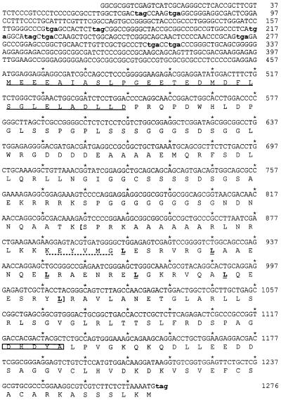
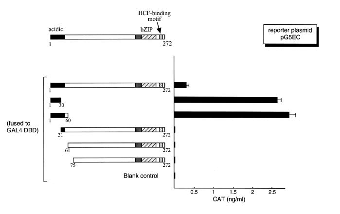
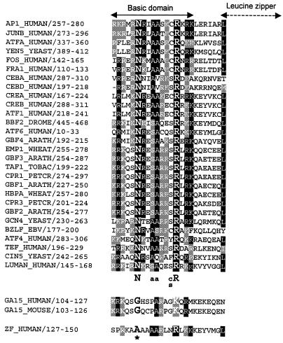
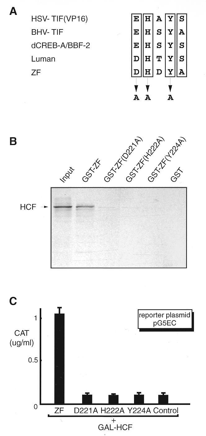
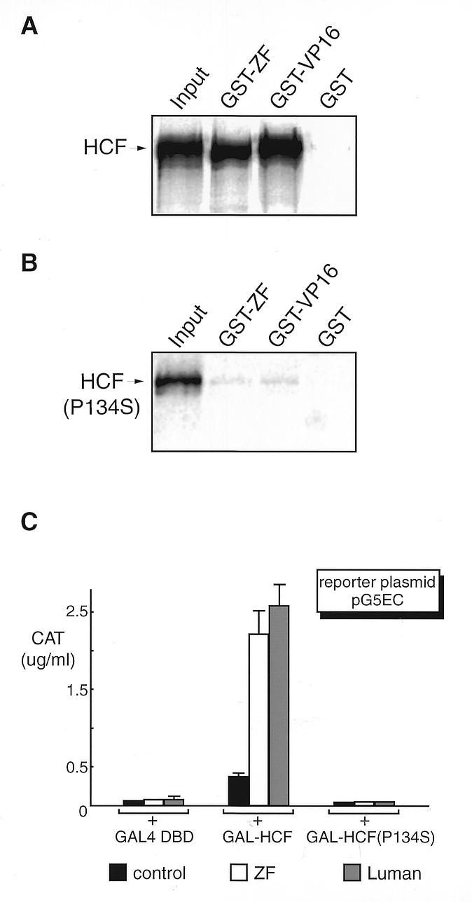
Host cell factor (HCF, C1, VCAF or CFF) is a cellular protein that is required for transcription activation of herpes simplex virus (HSV) immediate-early (IE) genes by the virion protein VP16. The biological function of HCF remains unclear. Recently we identified a cellular transcription activator, Luman. As with VP16, the transactivation function of Luman is also regulated by HCF. Here we report a second human protein, Zhangfei (ZF) that interacts with HCF in a fashion similar to Luman and VP16. Although ZF shares no significant sequence homology with Luman, the two proteins have some structural similarities. These include: a basic domain-leucine zipper (bZIP) region, an acidic activation domain and a consensus HCF-binding motif. Unlike Luman, or most other bZIP proteins, ZF by itself did not appear to bind consensus bZIP-binding sites. It was also unable to activate promoters containing these response elements. Although in transient expression assays ectopically expressed ZF was unable to block transactivation by VP16 of a HSV IE promoter, ZF could prevent the expression of several HSV proteins in cells infected with the virus. The ability of ZF to block the synthesis of the HSV IE protein ICP0 relied on its binding to HCF, since a mutant of ZF that was unable to bind HCF was also unable to prevent viral IE protein expression.


Figures

Figure 1
Figure 1
The nucleotide sequence of the ZF ORF and the known 5′-UTR. The identified cDNA contains only one ORF larger than 200 nt, with multiple upstream stop codons (lower case and bold) in all three reading frames. The ZF protein encoded by this ORF is 272 amino acids long with a predicted molecular mass of 29 kDa. This protein contains a bZIP region (in brackets) with six perfect leucine heptad repeats (bold and underlined). A conserved six amino acid spacer (dotted underlined) separating the basic domain and leucine zipper is also present. In the N-terminal region is an acidic domain (underlined), rich in negatively charged amino acids. Close to the C-terminal end it has a HCF-binding motif (boxed). The 1676 bp 3′-UTR containing multiple polyadenylation sites is not shown.
Figure 2
Figure 2
The N-terminal region of ZF, rich in negatively charged amino acids, is an activation domain. On the left is the schematic representation of the structure of the ZF protein. The numbers indicate the positions of the amino acid. ZF and its deletion mutants were fused to the GAL4 DNA-binding domain. The same amount (0.5 µg) of each plasmid was introduced into COS7 cells along with the reporter plasmid pG5EC (0.5 µg), which has five copies of the GAL4 UAS in the promoter region linked to the CAT gene. The parental vector expressing only the GAL4 DBD, pM1, was used as the blank control. The CAT activity was measured by ELISA 48 h post-transfection.
Figure 3
Figure 3
A sequence alignment of basic regions of 30 representative bZIP proteins and ZF. The numbers following the names of the gene locus indicate the location of the segment in the encoded protein. All bZIP proteins, except GA15 (or CHOP, GADD153 or DDIT3) and ZF, share a consensus quintet sequence, NXXAAXX(C/S)R (X stands for any amino acid), which is essential for DNA binding. ZF lacks the absolutely conserved residue N, which is critical for protein conformation; the amino portion of the basic region bears little resemblance to other bZIP factors.
Figure 4
Figure 4
Mutagenesis study of the HCF-binding motif in ZF. (A) The consensus sequence, [D/E]HXY[S/A], found in VP16 and Luman as well as their homologs is proved to be a HCF-binding motif. To test whether this motif in ZF is also involved in HCF binding, we mutated two absolutely conserved residues, H222 and Y224, and the negatively charged residue D221 by site-directed mutagenesis. (B) GST pull-down assay. GST and all GST fusion proteins were produced in E.coli strain BL21(DE3), coupled to glutathione–Sepharose beads. After incubation with [35S]HCF, the beads were washed and analyzed by 10% SDS–PAGE. The lane labeled input represents one-tenth of the [35S]HCF incubation mixture. (C) Mammalian two-hybrid assay. Plasmids pcZF, pcZF(D221A), pcZF(H222A) and pcZF(Y224A) were introduced into COS7 cells separately with reporter pG5EC and the plasmid expressing GAL–HCF. An aliquot of 0.5 µg DNA was used for each plasmid. Plasmid pcDNA3 was used as a control. The CAT activity was measured by ELISA 48 h post-transfection.
Figure 5
Figure 5
Like VP16, ZF binds to HCF but not to the mutant HCF(P134S), as determined by GST pull-down assay (A and B) and by mammalian two-hybrid assay (C). GST and GST fusion proteins of ZF and VP16 were produced in E.coli strain BL21(DE3), coupled to Sepharose beads. HCF was labeled with [35S]methionine by in vitro transcription and translation in a rabbit reticulocyte system (TnT; Promega). The protein-bound beads were incubated with an equivalent amount of 35S-labeled HCF (A) or HCF(P134S) (B). After extensive washing, proteins were eluted, fractionated by 10% SDS–PAGE and visualized by autoradiography. The first lane on the figure represents one-tenth of the input. Plasmids expressing GAL–HCF, GAL–HCF(P134) and GAL DBD alone (C) were co-transfected into COS7 cells with either pcZF, pcLuman or their parental vector pcDNA3 (0.5 µg each), with pG5EC as the reporter. The CAT activity was measured 48 h post-transfection.
Figure 6
Figure 6
ZF prevents HSV ICP0 expression in virus-infected cells in a HCF-dependent manner. Vero cells were transfected with plasmids expressing either ZF (AC) or ZF Y224A (DF). The cells were then infected with HSV-1 at a m.o.i. of 10 p.f.u./cell. Six hours after infection the cells were fixed and stained simultaneously for both ZF (Alexa546, red) and ICP0 (Alexa488, green). The cells were then visualized in a fluorescent microscope using either a 546 (A and D) or 450–490 nm filter (B and E). The red and green images were combined (C and F) using Northern Eclipse 5.0 software. In (E) the arrowheads indicate ICP0 stained nuclei that also stain for ZF Y224A.
Figure 7
Figure 7
Northern blot analysis of ZF. MTN northern blots (Clontech) containing poly(A)+ mRNA from adult and fetal human tissues were hybridized with 32P-labeled full-length ZF cDNA. Molecular size standards (in kilobases) and tissues of origin are indicated. The same blots were hybridized with the β-actin probe and used as a loading reference. Note that the human β-actin cDNA probe cross-hybridizes to muscle-type actin in heart and skeletal muscle.

References

    1. O’Hare P. (1993) Semin. Virol., 4, 145–155.
    1. Thompson C. and McKnight,S. (1992) Trends Genet., 8, 232–236.
    1. Kristie T.M. and Roizman,B. (1987) Proc. Natl Acad. Sci. USA, 84, 71–75. - PMC - PubMed
    1. Marsden H.S., Campbell,M.E., Haarr,L., Frame,M.C., Parris,D.S., Murphy,M., Hope,R.G., Muller,M.T. and Preston,C.M. (1987) J. Virol., 61, 2428–2437. - PMC - PubMed
    1. McKnight J.L., Kristie,T.M. and Roizman,B. (1987) Proc. Natl Acad. Sci. USA, 84, 7061–7065. - PMC - PubMed

Publication types

MeSH terms