. 2000 Jul 1;28(13):2481-93.
doi: 10.1093/nar/28.13.2481.

Structure-based predictions of Rad1, Rad9, Hus1 and Rad17 participation in sliding clamp and clamp-loading complexes

Affiliations

Structure-based predictions of Rad1, Rad9, Hus1 and Rad17 participation in sliding clamp and clamp-loading complexes

C Venclovas et al. Nucleic Acids Res. .

Abstract

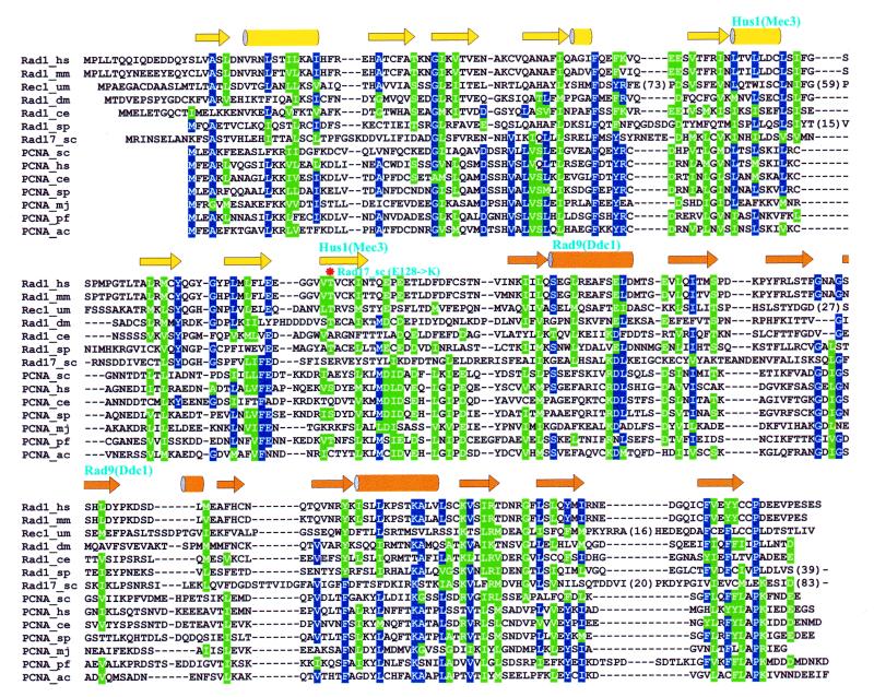
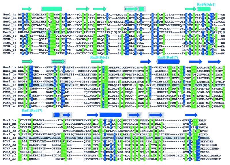
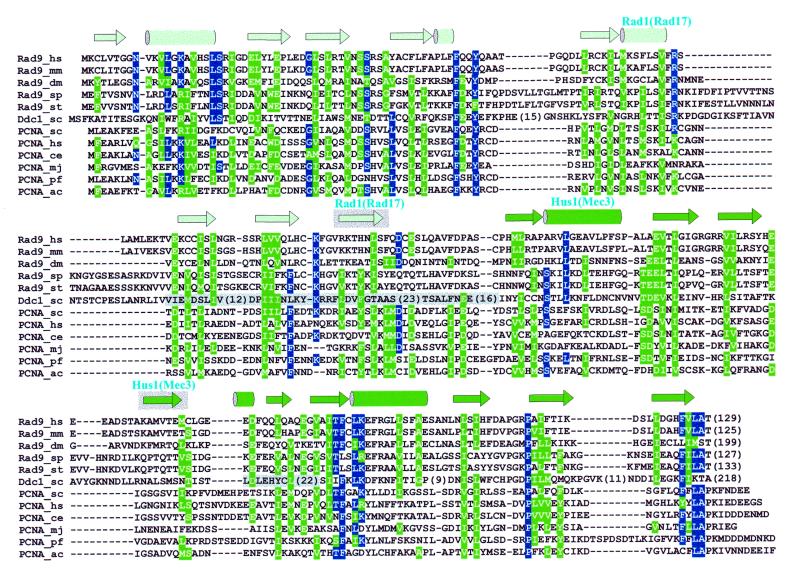
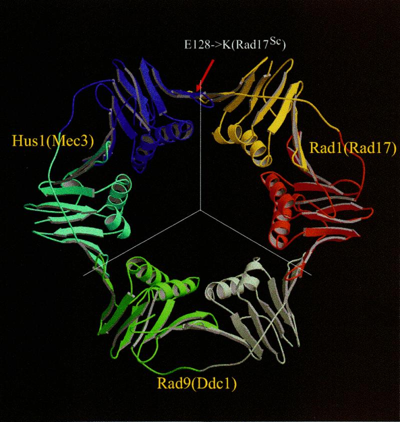
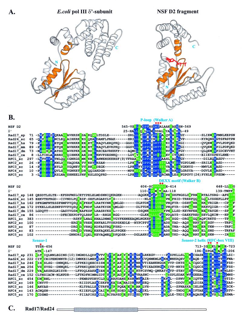
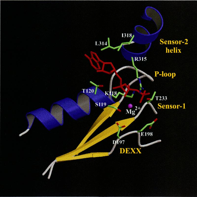
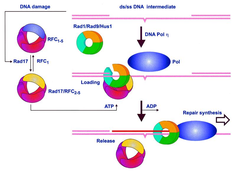
The repair of damaged DNA is coupled to the completion of DNA replication by several cell cycle checkpoint proteins, including, for example, in fission yeast Rad1(Sp), Hus1(Sp), Rad9(Sp) and Rad17(Sp). We have found that these four proteins are conserved with protein sequences throughout eukaryotic evolution. Using computational techniques, including fold recognition, comparative modeling and generalized sequence profiles, we have made high confidence structure predictions for the each of the Rad1, Hus1 and Rad9 protein families (Rad17(Sc), Mec3(Sc) and Ddc1(Sc) in budding yeast, respectively). Each of these families was found to share a common protein fold with that of PCNA, the sliding clamp protein that tethers DNA polymerase to its template. We used previously reported genetic and biochemical data for these proteins from yeast and human cells to predict a heterotrimeric PCNA-like ring structure for the functional Rad1/Rad9/Hus1 complex and to determine their exact order within it. In addition, for each individual protein family, contact regions with neighbors within the PCNA-like ring were identified. Based on a molecular model for Rad17(Sp), we concluded that members of this family, similar to the subunits of the RFC clamp-loading complex, are capable of coupling ATP binding with conformational changes required to load a sliding clamp onto DNA. This model substantiates previous findings regarding the behavior of Rad17 family proteins upon DNA damage and within the RFC complex of clamp-loading proteins.


Figures

Figure 1
Figure 1
Sequence–structure alignments of Rad checkpoint proteins with PCNA: (A) Rad1–PCNA, (B) Hus1–PCNA and (C) Rad9–PCNA. Residues conserved in sequences of more than a single family are colored blue (identical) and green (similar). Secondary structure of yeast PCNA is shown by arrows (strands) and cylinders (helices) for each alignment with different coloring representing two structural domains in a PCNA monomer. Regions of Rad1, Hus1 and Rad9 proteins involved in forming interfaces between monomers in the heterotrimeric ring structure (Fig. 2) are denoted by names of the interacting partners above the secondary structure elements. In the Rad1–PCNA alignment (A) the E128K mutation preventing formation of the Rad17Sc–Mec3Sc complex is also indicated (*). A shaded background for some secondary structure elements indicates that the sequence–structure alignment in corresponding regions is less confident. Shading in Mec3Sc (B) and Ddc1Sc (C) sequences mark regions of uncertainty in their alignment with, respectively, the Hus1 and Rad9 families. Long insertions and C-terminal regions extending beyond the PCNA fold were removed to preserve space and numbers in parentheses indicate the lengths of removed fragments.
Figure 1
Figure 1
Sequence–structure alignments of Rad checkpoint proteins with PCNA: (A) Rad1–PCNA, (B) Hus1–PCNA and (C) Rad9–PCNA. Residues conserved in sequences of more than a single family are colored blue (identical) and green (similar). Secondary structure of yeast PCNA is shown by arrows (strands) and cylinders (helices) for each alignment with different coloring representing two structural domains in a PCNA monomer. Regions of Rad1, Hus1 and Rad9 proteins involved in forming interfaces between monomers in the heterotrimeric ring structure (Fig. 2) are denoted by names of the interacting partners above the secondary structure elements. In the Rad1–PCNA alignment (A) the E128K mutation preventing formation of the Rad17Sc–Mec3Sc complex is also indicated (*). A shaded background for some secondary structure elements indicates that the sequence–structure alignment in corresponding regions is less confident. Shading in Mec3Sc (B) and Ddc1Sc (C) sequences mark regions of uncertainty in their alignment with, respectively, the Hus1 and Rad9 families. Long insertions and C-terminal regions extending beyond the PCNA fold were removed to preserve space and numbers in parentheses indicate the lengths of removed fragments.
Figure 2
Figure 2
Model of the Rad1/Rad9/Hus1 complex based on the PCNA structure. Line segments indicate boundaries between individual monomers. Coloring corresponds to that used in sequence–structure alignments in Figure 1. The red arrow points to a residue position corresponding to a point mutation in Rad17Sc that prevents complex formation with Mec3Sc. This and other structural figures were prepared with Molscript (53) and Raster3D (54).
Figure 3
Figure 3
(A) Comparison of 3D structures of the δ′ subunit of E.coli polymerase III and the NSF D2 fragment. Equivalent structural motifs involved in forming the nucleotide-binding site that could also be unambiguously aligned with the Rad17 and RFC families are colored gold. The ATP analog bound to NSF D2 is shown in red. (B) Alignment of the Rad17 family and all five yeast RFC subunits with the structural fragments of both δ′ and NSF D2, colored gold in (A), subsequently used to construct the model for Rad17Sp given in Figure 4. Residues conserved in more than half of the sequences are colored blue (identical) and green (similar). Red dots indicate Rad17Sp nucleotide-binding site residues shown with their side chains in Figure 4. Numbers indicate positions of residues in sequences. (C) Location of the Rad17 (Rad24Sc) region (gray rectangle) aligned in (B), found to be common to the AAA+ class (39).
Figure 4
Figure 4
Model of the Rad17Sp nucleotide-binding site. Side chains considered to be important for either binding ATP (red) or coordinating the magnesium ion (purple sphere) are shown for the same residues as those labeled in Figure 3B.
Figure 5
Figure 5
Proposed mechanism of Rad1, Rad9, Hus1 and Rad17 action in response to DNA damage. The DNA intermediate contains an unrepaired damaged site, such as a pyrimidine dimer (denoted by a bulge in the lower strand), that initiates translocation of Rad17 throughout the nucleus. Conversion of RFC1–5 to Rad17/RFC2–5 is required for ATP-dependent loading of the Rad1/Rad9/Hus1 heterotrimeric ring onto DNA. Repair synthesis can then proceed by (for example) a lesion bypass polymerase that utilizes the heterotrimeric ring for more efficient and/or accurate synthesis. See text for a more detailed description and references.

References

    1. Longhese M.P., Foiani,M., Muzi-Falconi,M., Lucchini,G. and Plevani,P. (1998) EMBO J., 17, 5525–5528. - PMC - PubMed
    1. Weinert T. (1998) Curr. Opin. Genet. Dev., 8, 185–193. - PubMed
    1. Huberman J.A. (1999) Prog. Nucleic Acid Res. Mol. Biol., 62, 369–395. - PubMed
    1. Caspari T., Dahlen,M., Kanter-Smoler,G., Lindsay,H.D., Hofmann,K., Papadimitriou,K., Sunnerhagen,P. and Carr,A.M. (2000) Mol. Cell. Biol., 20, 1254–1262. - PMC - PubMed
    1. Green C.M., Erdjument-Bromage,H., Tempst,P. and Lowndes,N.F. (2000) Curr. Biol., 10, 39–42. - PubMed

Publication types

MeSH terms