. 2000 Jul;66(7):2748-58.
doi: 10.1128/AEM.66.7.2748-2758.2000.

The secondary endosymbiotic bacterium of the pea aphid Acyrthosiphon pisum (Insecta: homoptera)

Affiliations

The secondary endosymbiotic bacterium of the pea aphid Acyrthosiphon pisum (Insecta: homoptera)

T Fukatsu et al. Appl Environ Microbiol. 2000 Jul.

Abstract

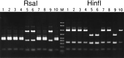
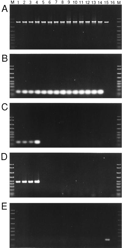
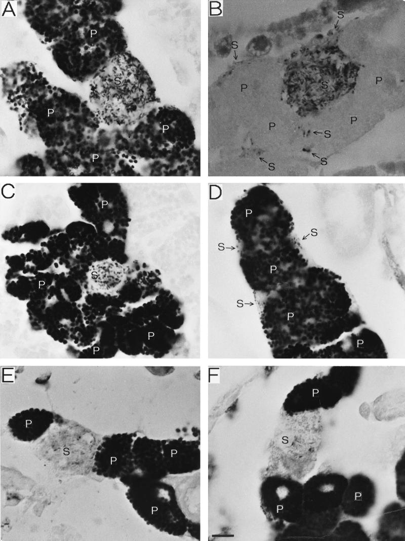
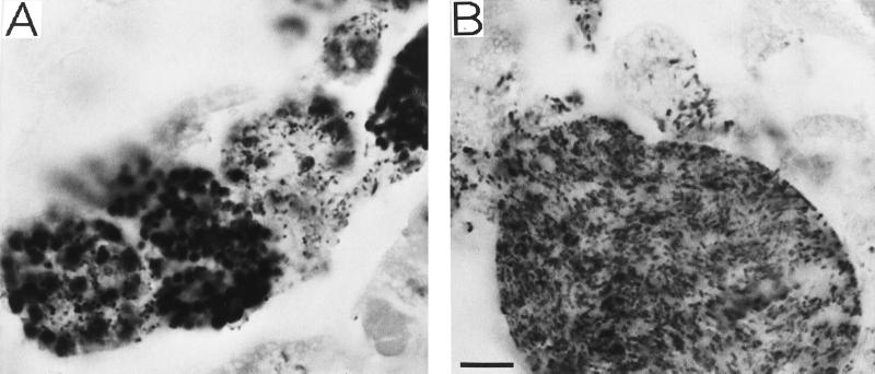
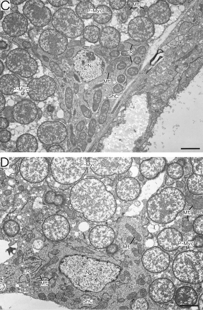
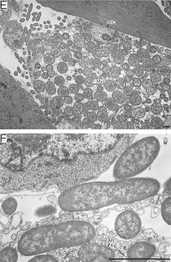
The secondary intracellular symbiotic bacterium (S-symbiont) of the pea aphid Acyrthosiphon pisum was investigated to determine its prevalence among strains, its phylogenetic position, its localization in the host insect, its ultrastructure, and the cytology of the endosymbiotic system. A total of 14 aphid strains were examined, and the S-symbiont was detected in 4 Japanese strains by diagnostic PCR. Two types of eubacterial 16S ribosomal DNA sequences were identified in disymbiotic strains; one of these types was obtained from the primary symbiont Buchnera sp., and the other was obtained from the S-symbiont. In situ hybridization and electron microscopy revealed that the S-symbiont was localized not only in the sheath cells but also in a novel type of cells, the secondary mycetocytes (S-mycetocytes), which have not been found previously in A. pisum. The size and shape of the S-symbiont cells were different when we compared the symbionts in the sheath cells and the symbionts in the S-mycetocytes, indicating that the S-symbiont is pleomorphic under different endosymbiotic conditions. Light microscopy, electron microscopy, and diagnostic PCR revealed unequivocally that the hemocoel is also a normal location for the S-symbiont. Occasional disordered localization of S-symbionts was also observed in adult aphids, suggesting that there has been imperfect host-symbiont coadaptation over the short history of coevolution of these organisms.


Figures

FIG. 1
FIG. 1
RFLP analysis of bacterial 16S rDNA amplified and cloned from the total DNA of A. pisum IS. Lanes 1 through 10 contained cloned 16S rDNA fragments digested by RsaI (left) or HinfI (right) and resolved in a 2% agarose gel. Lanes 1 through 4, 7, 8, and 10, clones containing the P-symbiont sequence; lanes 5, 6, and 9, clones containing the S-symbiont sequence. Lane M contained DNA size markers (2,000, 1,500, 1,000, 700, 500, 400, 300, 200, and 100 bp, from top to bottom).
FIG. 2
FIG. 2
Diagnostic PCR detection of endosymbionts in A. pisum strains. (A) Universal detection of eubacterial endosymbionts with primers 16SA1 and 16SB1. (B) Specific detection of the P-symbiont with primers 16SA1 and ApisP. (C) Specific detection of the S-symbiont with primers 16SA1 and ApisS. (D) Specific detection of the S-symbiont with primers 16SA1 and PASScmp. (E) Specific detection of Rickettsia spp. with primers 16SA1 and Rick16SR. Lane 1, strain IS; lane 2, TKC93; lane 3, MR88; lane 4, HG99; lane 5, EF99; lane 6, SM; lane 7, AIST99; lane 8, LL01; lane 9, LL02; lane 10, LL04; lane 11, LL05; lane 12, LC06; lane 13, LF08; lane 14, LC09; lane 15, bruchid beetle Kytorhinus sharpianus containing Rickettsia sp. (24); lane 16, no-template control; lane M, DNA size markers (2,000, 1,500, 1,000, 700, 500, 400, 300, 200, and 100 bp, from top to bottom). Although the results for only one individual of each strain are shown, the reproducibility of the results was confirmed by examining more than 12 individuals of each strain.
FIG. 3
FIG. 3
In situ hybridization of the P- and S-symbionts of A. pisum. (A) Mycetome of a strain IS embryo probed with BIO-EUB338. Tubular S-symbionts in a S-mycetocyte and globular P-symbionts in many P-mycetocytes are present. (B) Mycetome of a strain IS embryo probed with BIO-PASScmp. Tubular S-symbionts in a S-mycetocyte and small S-symbionts in sheath cells are specifically visualized in the mycetome. (C) Mycetome of a strain IS embryo probed with BIO-EUB338. In this S-mycetocyte, the S-symbionts are short rods. (D) Mycetome of a strain IS embryo probed with BIO-EUB338. Sheath cells containing small S-symbionts are associated with the P-mycetocytes. (E) Mycetome of a strain MR88 embryo probed with BIO-EUB338. A S-mycetocyte harboring tubular S-symbionts is located between P-mycetocytes. (F) Mycetome of a strain TKC93 embryo probed with BIO-EUB338. The same disymbiotic organization is observed. Bar = 10 μm. The arrows indicate the locations of sheath cells. Abbreviations: P, P-symbiont: S, S-symbiont.
FIG. 4
FIG. 4
Disordered localization of the S-symbiont found in several unwinged adults of A. pisum IS. (A) Embryonic P-mycetocytes infected by S-symbionts. Abnormally, the P- and S-symbionts coexist in several cells. (B) Maternal P-mycetocyte filled with S-symbionts. The P-symbionts are almost completely replaced by S-symbionts. The symbionts were visualized by in situ hybridization with probe BIO-EUB338. Bar = 10 μm.
FIG. 5
FIG. 5
Electron microscopy of the endosymbiosis in A. pisum embryos. (A through C, E, and F) Strain IS containing both P- and S-symbionts. (D) Strain SM lacking the S-symbiont. (A) Strain IS sheath cell located between large P-mycetocytes. Small rod-shaped S-symbionts are located intracellularly. (B) Strain IS S-mycetocyte harboring many tubular S-symbionts. The S-symbionts are apparently larger than the S-symbionts in sheath cells. The cytoplasm of the S-mycetocyte is highly vacuolated. (C) Magnified image of a strain IS sheath cell containing S-symbionts, mitochondria, and endoplasmic reticulum. (D) Magnified image of a sheath cell of a strain SM aphid without the S-symbiont. The sheath cell contains developed mitochondria and endoplasmic reticulum but no S-symbionts. (E) Extracellular S-symbionts associated with strain IS sheath cells on the periphery of a mycetome. (F) Magnified image of S-symbionts on the periphery of a strain IS sheath cell. Bars = 2 μm. Abbreviations: ER, endoplasmic reticulum; Mt, mitochondrion; N, nucleus; P-Myc, primary mycetocyte; ShC, sheath cell; S-Myc, secondary mycetocyte.
FIG. 5
FIG. 5
Electron microscopy of the endosymbiosis in A. pisum embryos. (A through C, E, and F) Strain IS containing both P- and S-symbionts. (D) Strain SM lacking the S-symbiont. (A) Strain IS sheath cell located between large P-mycetocytes. Small rod-shaped S-symbionts are located intracellularly. (B) Strain IS S-mycetocyte harboring many tubular S-symbionts. The S-symbionts are apparently larger than the S-symbionts in sheath cells. The cytoplasm of the S-mycetocyte is highly vacuolated. (C) Magnified image of a strain IS sheath cell containing S-symbionts, mitochondria, and endoplasmic reticulum. (D) Magnified image of a sheath cell of a strain SM aphid without the S-symbiont. The sheath cell contains developed mitochondria and endoplasmic reticulum but no S-symbionts. (E) Extracellular S-symbionts associated with strain IS sheath cells on the periphery of a mycetome. (F) Magnified image of S-symbionts on the periphery of a strain IS sheath cell. Bars = 2 μm. Abbreviations: ER, endoplasmic reticulum; Mt, mitochondrion; N, nucleus; P-Myc, primary mycetocyte; ShC, sheath cell; S-Myc, secondary mycetocyte.

References

    1. Adachi J, Hasegawa M. MOLPHY version 2.3: programs for molecular phylogenetics based on maximum likelihood. Comput Sci Monogr Inst Stat Math Tokyo. 1996;28:1–150.
    1. Amann R I, Krumholz L, Stahl D A. Fluorescent-oligonucleotide probing of whole cells for determinative, phylogenetic, and environmental studies in microbiology. J Bacteriol. 1990;172:762–770. - PMC - PubMed
    1. Baumann P, Moran N A. Non-cultivable microorganisms from symbiotic associations of insects and other hosts. Antonie Leeuwenhoek. 1997;72:38–48. - PubMed
    1. Baumann P, Baumann L, Lai C-Y, Rouhbakhsh D, Moran N A, Clark M A. Genetics, physiology and evolutionary relationships of the genus Buchnera: intracellular symbionts of aphids. Annu Rev Microbiol. 1995;49:55–94. - PubMed
    1. Blackman R L, Eastop V F. Aphids on the world's trees. Wallingford, United Kingdom: CAB International; 1994.

Publication types

LinkOut - more resources