. 2000 Jul 1;526 Pt 1(Pt 1):157-65.
doi: 10.1111/j.1469-7793.2000.t01-1-00157.x.

Effect of pulmonary C-fibre afferent stimulation on cardiac vagal neurones in the nucleus ambiguus in anaesthetized cats

Affiliations

Effect of pulmonary C-fibre afferent stimulation on cardiac vagal neurones in the nucleus ambiguus in anaesthetized cats

Y Wang et al. J Physiol. .

Abstract

It has been demonstrated previously that the vagal bradycardia evoked by activation of pulmonary C-fibres is not respiratory modulated. Experiments were carried out in alpha-chloralose anaesthetized cats to determine if these cardiac vagal preganglionic neurones (CVPNs) in the nucleus ambiguus (NA), which have respiratory modulated activity, can be activated when pulmonary C-fibre afferents are stimulated by right atrial injections of phenylbiguanide (PBG). Eleven CVPNs with B-fibre axons in the right cardiac vagal branches were identified and found to be localized within or ventrolateral to the nucleus ambiguus. Ionophoretic application of a high current of dl-homocysteic acid (DLH) induced a vagally mediated bradycardia and hypotension in six of eight sites from which CVPNs were recorded. The activity of B-fibre CVPNs, whether spontaneous (n = 4) or induced by ionophoresis of DLH (n = 7) was respiratory modulated, firing perferentially during post-inspiration and stage 2 expiration. This activity also correlated with the rising phase of the arterial blood pressure wave consistent with these CVPNs receiving an arterial baroreceptor input. Right atrial injections of PBG excited nine of eleven CVPNs tested. In eight of these activated neurones the onset latency of the excitation was within the pulmonary circulation time, consistent with being activated only by pulmonary C-fibre afferents. In two neurones the PBG-evoked excitation still occurred when central inspiratory drive was inhibited, as indicated by the disappearance of phrenic nerve activity. In conclusion, B-fibre respiratory modulated CVPNs can be activated following stimulation of pulmonary C-fibre afferents.


Figures

Figure 1
Figure 1. Identification of a B-fibre cardiac vagal preganglionic neurone in the nucleus ambiguus
A, traces showing a cardiac vagal preganglionic neurone antidromically activated (latency, 15 ms) by stimulating the right cardiac branch (200 μA, 1 ms, 0.5 Hz). Aa, five consecutive sweeps superimposed to show the constant latency of the evoked spike; Ab, three consecutive sweeps showing that the evoked spike (see top and bottom trace) was cancelled by the spontaneous spike (see middle trace). The • indicates the stimulus artefacts. B, histograms of the activity (with DLH at 20 nA) of the same CVPN as in A triggered by integrated phrenic nerve activity (Int-phre; 50 ms bin width; top panel), the R-wave of the ECG (10 ms bin width; middle panel) and by tracheal pressure (10 ms bin width; lower panel). Above the histograms is an average of integrated phrenic activity, ECG triggered arterial blood pressure (BP) and the tracheal pressure (TP) wave, respectively. The number of sweeps on top of each panel refers to both the average and to the histogram.
Figure 2
Figure 2. Pontamine Sky Blue marked locations of recordings from the medulla
The position of six B-fibre (•) cardiac vagal preganglionic neurones from which recordings were made are shown on five standard sections of the medulla taken from -1 to +2.5 mm caudal to rostral at the level of the obex. Abbreviations: NA, nucleus ambiguus; NTS, nucleus tractus solitarius; X, dorsal vagal nucleus; XII, hypoglossal nucleus; AP, area postrema.
Figure 3
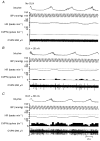
Figure 3. Traces showing the effects of ionophoretic application of DLH onto a CVPN with a B-fibre axon
Anaesthetized cat pretreated with atenolol (1 mg kg−1, i.v.). Records from top downwards: Int-phre, integrated phrenic nerve activity; BP, arterial blood pressure (mmHg); HR, heart rate (beats min−1) and rate histogram (0.5 s bin) and recording of the activity of a B-fibre CVPN. A, ongoing activity, no DLH; B and C, activity in the presence of 20 nA DLH and 60 nA DLH, respectively. Note the heart rate fell as CVPN discharge increased (the dotted lines on the heart rate traces represent the mean heart rate in control without DLH, 150 beats min−1) and even at a high level of excitation the activity was still respiratory modulated (C).
Figure 4
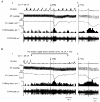
Figure 4. Traces comparing the effect of right atrial injections (at the point marked by arrow) of PBG (20 μg kg−1) on a CVPN with a B-fibre axon in the presence (A) and absence (B) of central respiratory drive
Anaesthetized cat pretreated with atenolol (1 mg kg−1, i.v.). Records from top downwards: Int-phre, integrated phrenic nerve activity; BP, arterial blood pressure (mmHg); HR, heart rate (beats min−1) and CVPN rate histogram (0.5 s bin) and on-going activity. The 5 s window following the PBG injection (see Methods) is shown by the two vertical dotted lines. Ab and Bb are expanded traces after the PBG injections shown in Aa and Ba. Note in B: (1) low intensity electrical stimulation of the pulmonary vagal branch inhibits central respiratory drive, as indicated by the lack of phrenic nerve activity, and (2) the increase in neuronal activity during this stimulation is due to the inhibition of central respiratory drive.
Figure 5
Figure 5. Synaptic input from vagal branches to two different CVPNs with B-fibre axons
Two sets of traces containing ten consecutive sweeps each showing the effect of stimulation (•) of the pulmonary branch (200 μA, 1 Hz, 1 ms) (A), which evoked both a short and a long latency excitatory input (Note: in the 6th trace (*) the spontaneous spike did not cancel the short latency input from the pulmonary nerve), and the cardiac branch (150 μA, 0.5 Hz, 1 ms) (B), which evoked a short latency antidromic spike (○) and a long latency synaptic input.

References

    1. Bennett JA, Goodchild CS, Kidd C, McWilliam PN. Neurones in the brain stem of the cat excited by vagal afferent fibres from the heart and lungs. The Journal of Physiology. 1985;369:1–15. - PMC - PubMed
    1. Berman AL. The Brainstem of the Cat. Madison, WI, USA: University of Wisconsin; 1968.
    1. Coleridge JCG, Coleridge HM. Handbook of Physiology, section 2, The Cardiovascular System, The Heart. Vol. 1. Bethesda, MD, USA: American Physiological Society; 1979. Chemoreflex regulation of the heart; pp. 653–676.
    1. Coleridge JCG, Coleridge HM. Afferent vagal C-fibre innervation of the lungs and airways and its function significance. Reviews of Physiology, Biochemistry and Pharmacology. 1984;99:1–110. - PubMed
    1. Daly MdeB. Some reflex cardioinhibitory responses in the cat and their modulation by central inspiratory neuronal activity. The Journal of Physiology. 1991;439:559–577. - PMC - PubMed

Publication types

MeSH terms

LinkOut - more resources