. 2000 Jul;106(1):37-45.
doi: 10.1172/JCI9102.

Antiviral treatment normalizes neurophysiological but not movement abnormalities in simian immunodeficiency virus-infected monkeys

Affiliations

Antiviral treatment normalizes neurophysiological but not movement abnormalities in simian immunodeficiency virus-infected monkeys

H S Fox et al. J Clin Invest. 2000 Jul.

Abstract

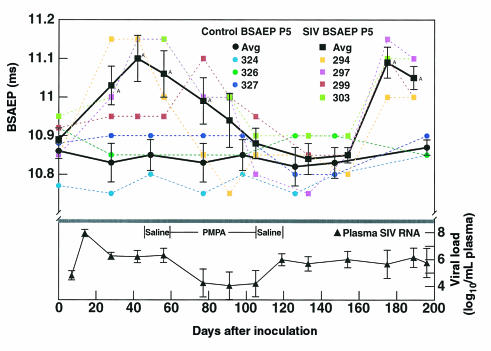
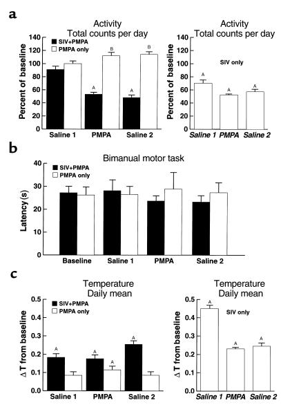
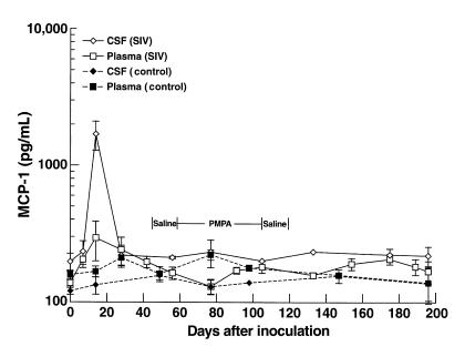
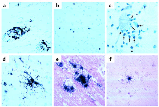
Simian immunodeficiency virus (SIV) infection of rhesus monkeys provides an excellent model of the central nervous system (CNS) consequences of HIV infection. To discern the relationship between viral load and abnormalities induced in the CNS by the virus, we infected animals with SIV and later instituted antiviral treatment to lower peripheral viral load. Measurement of sensory-evoked potentials, assessing CNS neuronal circuitry, revealed delayed latencies after infection that could be reversed by lowering viral load. Cessation of treatment led to the reappearance of these abnormalities. In contrast, the decline in general motor activity induced by SIV infection was unaffected by antiviral treatment. An acute increase in the level of the chemokine monocyte chemoattractant protein-1 (MCP-1) was found in the cerebrospinal fluid (CSF) relative to plasma in the infected animals at the peak of acute viremia, likely contributing to an early influx of immune cells into the CNS. Examination of the brains of the infected animals after return of the electrophysiological abnormalities revealed diverse viral and inflammatory findings. Although some of the physiological abnormalities resulting from SIV infection can be at least temporarily reversed by lowering viral load, the viral-host interactions initiated by infection may result in long-lasting changes in CNS-mediated functions.


Figures

Figure 1
Figure 1
Effects of PMPA on SIV-induced evoked potential abnormalities. Top: BSAEP P5 latencies in groups of four SIV-infected and three uninfected control monkeys. The latency is indicated on the y-axis. SIV-infected animals are indicated by squares, control uninfected animals by circles. Black symbols are the group averages with error bars denoting SEM; colored symbols show data points for the individual animals. After infection, the evoked potential latency is significantly delayed 1–2 months after infection but becomes indistinguishable from preinoculation values after 1 month of PMPA treatment. When treatment is withdrawn, BSAEP latency values remain normal for approximately 2 months, then again become significantly delayed. AThe point differs from baseline. Bottom: Copy equivalents of SIV RNA in the plasma of the infected monkeys. Plasma viral load, indicated on the y-axis (log10 transformed, error bars indicate SEM), peaks at 14 days after inoculation, then reaches a steady-state level. Upon institution of PMPA therapy, viral load drops more than 100-fold but recovers to pretreatment levels upon cessation of therapy.
Figure 2
Figure 2
Effects of PMPA and SIV infection on movement and temperature. (a) Gross motor activity. Gross motor activity was normalized for each animal to its baseline, and data for each group were expressed as a percentage of baseline along the y-axis. The heights of the boxes indicate the group mean, and the error bars the SEM. Three time periods are delineated: saline injection before PMPA treatment (Saline 1); during PMPA treatment (PMPA); and saline injection after PMPA treatment (Saline 2). In the left panel, data from the SIV-infected (filled boxes) and uninfected (open boxes) animals in this study are shown. A decrease in gross motor activity followed SIV infection regardless of PMPA administration. In the right panel, data are included for comparison from the same period after viral inoculation (indicated in italics) in a previously studied untreated SIV-infected group (10). AThe point differed from baseline condition. BThe groups differed during this condition. (b) Fine motor coordination in the bimanual motor task. The latency to retrieve all 15 raisins is indicated on the y-axis (error bars represent the SEM) for the time points indicated as in a. Despite large changes in gross motor activity, there were no effects of either PMPA or SIV infection on fine motor control in the bimanual motor task. (c) Body temperature. Change in temperature (ΔT) from baseline, plotted on the y-axis with error bars indicating the SEM, was determined by subtracting the baseline mean from each daily mean temperature. The periods analyzed and the animal groups are as in a. An increase in body temperature followed SIV infection regardless of PMPA administration. In the uninfected animals, the injections resulted in a slight increase in body temperature, which was significantly above baseline during the PMPA treatment (left panel). AThe point differs from the baseline condition.
Figure 3
Figure 3
MCP-1 concentrations in the plasma and CSF of SIV-infected monkeys. The y-axis indicates the mean MCP-1 concentration in the CSF (open diamonds) and plasma (open squares) in the SIV-infected animals, and in the CSF (filled diamonds) and plasma (filled squares) over the time course of the experiment. The error bars indicate the SEM. An increased ratio of MCP-1 in the CSF relative to plasma (5.8-fold) was seen at 14 days after inoculation in the infected animals, coincident with the peak of viral load. All four infected animals exhibited higher levels of MCP-1 in the CSF than in plasma at this time point. At other time points, CSF levels ranged from 0.9 to 1.8 of those in plasma.
Figure 4
Figure 4
Histopathology in the CNS of PMPA-treated, SIV-infected monkeys. (a) Mac387 reactivity in perivascular macrophages in monkey 297. (b) LCA reactivity of infiltrating lymphocytes in monkey 294. (c) Granzyme B reactivity in infiltrating lymphocytes in monkey 303. Cells (arrows) expressing immunoreactive granules surround a neuron. (d) LN3-reactivity for HLA-DR in a cell with the morphological characteristics of a resident microglial cell (center). (e) In situ hybridization revealing cells producing SIV RNA in animal 297. (f) In situ hybridization revealing a cell producing SIV RNA in animal 294.

Comment in

  • HIV and HIV dementia.
    Kolson DL, González-Scarano F. Kolson DL, et al. J Clin Invest. 2000 Jul;106(1):11-3. doi: 10.1172/JCI10553. J Clin Invest. 2000. PMID: 10880043 Free PMC article. No abstract available.

References

    1. Price RW, et al. The brain in AIDS: central nervous system HIV-1 infection and AIDS dementia complex. Science. 1988;239:586–592. - PubMed
    1. Heaton RK, et al. Neuropsychological impairment in human immunodeficiency virus-infection: implications for employment. HNRC Group. HIV Neurobehavioral Research Center. Psychosom Med. 1994;56:8–17. - PubMed
    1. Michaels SH, Clark R, Kissinger P. Declining morbidity and mortality among patients with advanced human immunodeficiency virus infection. N Engl J Med. 1998;339:405–406. - PubMed
    1. Moore RD, Chaisson RE. Natural history of HIV infection in the era of combination antiretroviral therapy. AIDS. 1999;14:1933–1942. - PubMed
    1. Dore GJ, et al. Changes to AIDS dementia complex in the era of highly active antiretroviral therapy. AIDS. 1999;13:1249–1253. - PubMed

Publication types

MeSH terms

Substances