

Distribution and phenotype of Epstein-Barr virus-infected cells in inflammatory bowel disease
- PMID: 10880375
- PMCID: PMC1850210
- DOI: 10.1016/S0002-9440(10)64516-6
Distribution and phenotype of Epstein-Barr virus-infected cells in inflammatory bowel disease
Abstract
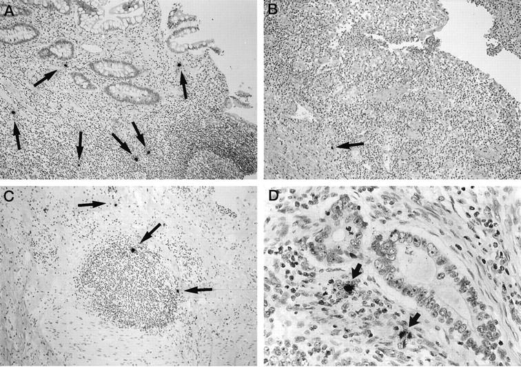
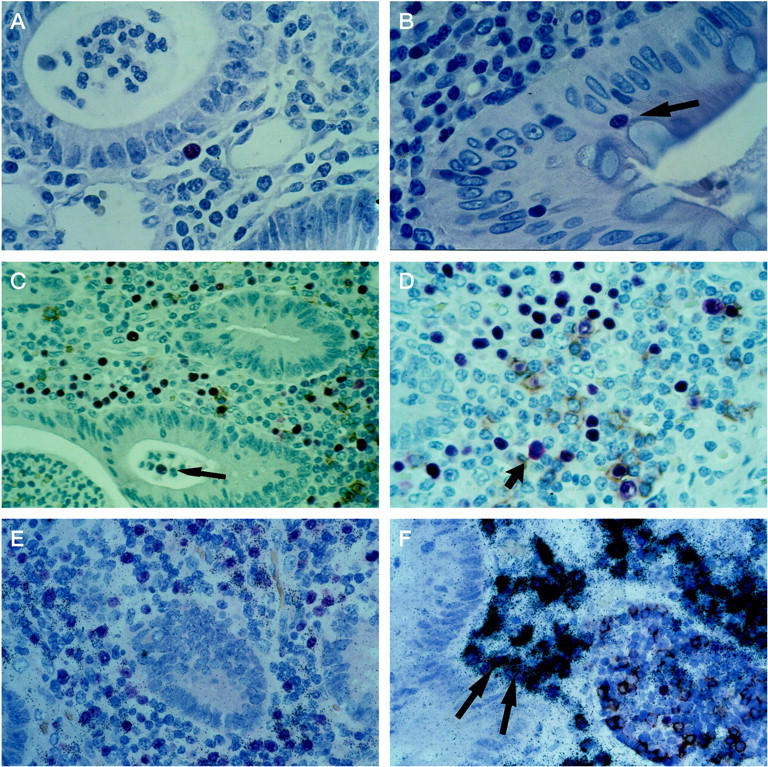
Little is known about Epstein-Barr virus (EBV) infection of colon mucosa, particularly in inflammatory bowel diseases. Crohn's disease and ulcerative colitis are thought to differ in T-helper lymphocyte composition and cytokine secretion patterns. Some of the implicated cytokines are growth factors for EBV-infected cells. We examined colon mucosa for differences in the distribution and phenotype of EBV-infected cells. Colon tissues with Crohn's disease (n = 31) or ulcerative colitis (n = 25) and controls (n = 60) were characterized by in situ hybridization and immunohistology for six EBV gene products as indicators of latent and replicative EBV infection. The cells were additionally phenotyped by combined detection of the EBV transcripts and B- or T-cell antigens. B lymphocytes predominated as the site of latent EBV infection in the colon and were most numerous in ulcerative colitis. In active ulcerative colitis, EBV-positive lymphocytes accumulated under and within the epithelium and displayed evidence for replicative infection. The patterns of mucosal EBV gene expression indicate local impairment of virus-specific T-cell responses in active ulcerative colitis. Detection of EBV may help to discriminate between active ulcerative colitis and other inflammatory bowel diseases. Colon mucosa is a potential site of EBV replication and may be relevant for EBV transmission.
Figures
Similar articles
-
Epstein-Barr virus (EBV) infection and expression of the interleukin-12 family member EBV-induced gene 3 (EBI3) in chronic inflammatory bowel disease.J Med Virol. 2004 Jul;73(3):432-8. doi: 10.1002/jmv.20109. J Med Virol. 2004. PMID: 15170639
-
Epstein-Barr virus infection of the colon with inflammatory bowel disease.Am J Gastroenterol. 1999 Jun;94(6):1582-6. doi: 10.1111/j.1572-0241.1999.01148.x. Am J Gastroenterol. 1999. PMID: 10364028
-
Epstein-Barr virus infection in colorectal neoplasms associated with inflammatory bowel disease: detection of the virus in lymphomas but not in adenocarcinomas.J Pathol. 2003 Oct;201(2):312-8. doi: 10.1002/path.1442. J Pathol. 2003. PMID: 14517849
-
In situ detection of Epstein-Barr virus and phenotype determination of EBV-infected cells.Methods Mol Biol. 2006;326:115-37. doi: 10.1385/1-59745-007-3:115. Methods Mol Biol. 2006. PMID: 16780197 Review.
-
Regulation and dysregulation of Epstein-Barr virus latency: implications for the development of autoimmune diseases.Autoimmunity. 2008 May;41(4):298-328. doi: 10.1080/08916930802024772. Autoimmunity. 2008. PMID: 18432410 Review.
Cited by
-
Epstein-barr virus-related diarrhea or exacerbation of inflammatory bowel disease: diagnostic dilemma.J Clin Microbiol. 2009 May;47(5):1588-90. doi: 10.1128/JCM.02477-08. Epub 2009 Mar 11. J Clin Microbiol. 2009. PMID: 19279175 Free PMC article.
-
Infection with murine gammaherpesvirus 68 exacerbates inflammatory bowel disease in IL-10-deficient mice.Inflamm Res. 2009 Dec;58(12):881-9. doi: 10.1007/s00011-009-0059-x. Epub 2009 Jun 21. Inflamm Res. 2009. PMID: 19544045
-
Epstein-Barr virus infection is common in inflamed gastrointestinal mucosa.Dig Dis Sci. 2012 Jul;57(7):1887-98. doi: 10.1007/s10620-012-2116-5. Epub 2012 Mar 13. Dig Dis Sci. 2012. PMID: 22410851 Free PMC article.
-
Drosophila melanogaster as a Model System to Assess the Effect of Epstein-Barr Virus DNA on Inflammatory Gut Diseases.Front Immunol. 2021 Mar 22;12:586930. doi: 10.3389/fimmu.2021.586930. eCollection 2021. Front Immunol. 2021. PMID: 33828545 Free PMC article.
-
Epstein-Barr virus infection in ulcerative colitis: a clinicopathologic study from a Chinese area.Therap Adv Gastroenterol. 2020 Aug 18;13:1756284820930124. doi: 10.1177/1756284820930124. eCollection 2020. Therap Adv Gastroenterol. 2020. PMID: 32913442 Free PMC article.
References
-
- Rickinson AB, Kieff E: Epstein-Barr virus. Fields Virology, vol. 2, ed 2. Edited by BN Fields, DM Knipe, PM Howley. Philadelphia, Lippincott-Raven, 1996, pp 2397–2446
-
- Anagnostopoulos I, Hummel M, Kreschel C, Stein H: Morphology, immunophenotype, and distribution of latently and/or productively Epstein-Barr virus-infected cells in acute infectious mononucleosis: implications for the interindividual infection route of Epstein-Barr virus. Blood 1995, 85:744-750 - PubMed
-
- Niedobitek G, Agathangellou A, Herbst H, Whitehead L, Wright DH, Young LS: Epstein-Barr virus (EBV) infection in infectious mononucleosis: virus latency, replication and phenotype of EBV-infected cells. J Pathol 1997, 182:151-159 - PubMed
-
- Herbst H, Foss HD, Samol J, Araujo I, Klotzbach H, Krause H, Agathanggelou A, Niedobitek G, Stein H: Frequent expression of interleukin-10 by Epstein-Barr virus-harboring tumor cells of Hodgkin’s disease. Blood 1996, 87:2918-2929 - PubMed
-
- Paul WE: Interleukin-4: a prototypic immunoregulatory lymphokine. Blood 1991, 77:1859-1870 - PubMed
Publication types
MeSH terms
Substances
LinkOut - more resources
Full Text Sources
Medical
