. 2000 Jul;157(1):135-44.
doi: 10.1016/S0002-9440(10)64525-7.

Overexpression of vascular endothelial growth factor (VEGF) in the retinal pigment epithelium leads to the development of choroidal neovascularization

Affiliations

Overexpression of vascular endothelial growth factor (VEGF) in the retinal pigment epithelium leads to the development of choroidal neovascularization

K Spilsbury et al. Am J Pathol. 2000 Jul.

Erratum in

  • Am J Pathol 2000 Oct;157(4):1413

Abstract

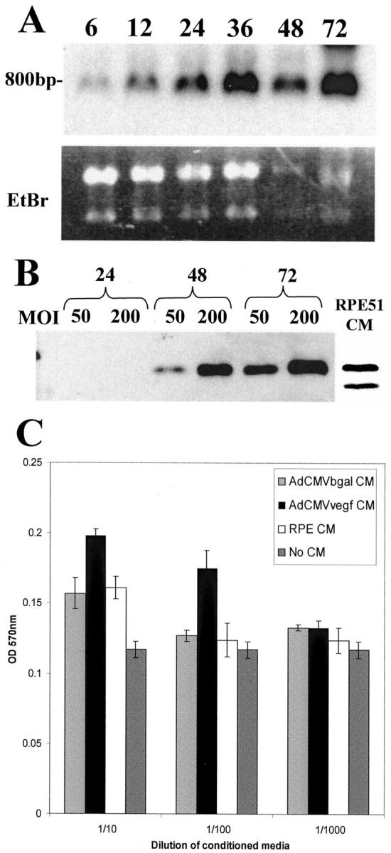
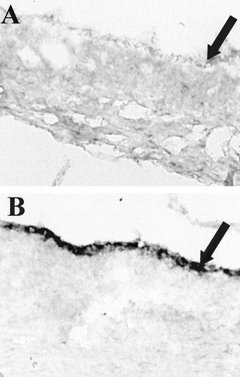
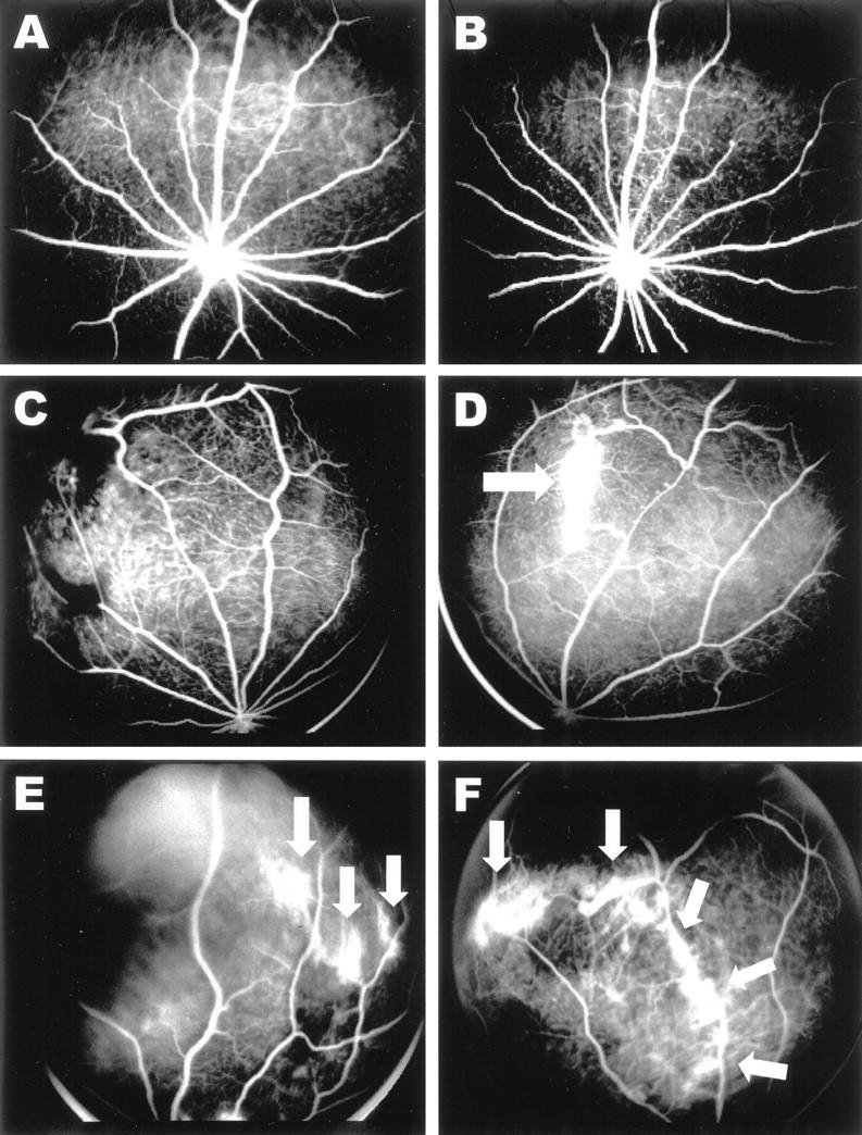
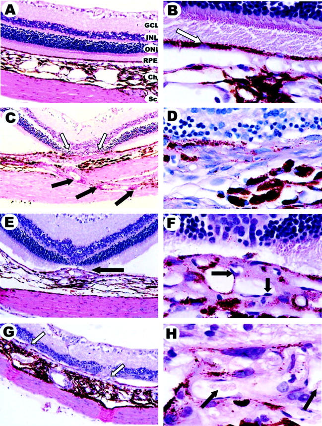
Vascular endothelial growth factor (VEGF) has been strongly implicated in the development of choroidal neovascularization found in age-related macular degeneration. Normally expressed in low levels, this study investigates whether the overexpression of VEGF in the retinal pigment epithelium is sufficient to cause choroidal neovascularization in the rat retina. A recombinant adenovirus vector expressing the rat VEGF(164) cDNA (AdCMV.VEGF) was constructed and injected into the subretinal space. The development of neovascularization was followed by fluorescein angiography, which indicates microvascular hyperpermeability of existing and/or newly forming blood vessels, and histology. VEGF mRNA was found to be overexpressed by retinal pigment epithelial cells and resulted in leaky blood vessels at 10 days postinjection, which was maintained for up to 31 days postinjection. By 80 days postinjection, new blood vessels had originated from the choriocapillaris, grown through the Bruch's membrane to the subretinal space, and disrupted the retinal pigment epithelium. This ultimately led to the formation of choroidal neovascular membranes and the death of overlying photoreceptor cells. By controlling the amount of virus delivered to the subretinal space, we were able to influence the severity and extent of the resulting choroidal neovascularization. These results show that even temporary overexpression of VEGF in retinal pigment epithelial cells is sufficient to induce choroidal neovascularization in the rat eye.


Figures

Figure 1.
Figure 1.
A: Northern blot analysis of rat VEGF164 mRNA produced from AdCMV.VEGF-transduced human RPE 51 cells harvested at 6, 12, 24, 36, 48, and 72 hours post-transduction. AdCMV.VEGF mRNA was approximately 800 bp in size. Ethidium bromide staining showed equality and integrity of RNA loading. B: Western blot analysis of rat VEGF164 produced from AdCMV.VEGF-transduced human RPE 51 cells. Conditioned medium from human RPE 51 cells transduced at a MOI of either 50 or 200 with AdCMV.VEGF harvested at 24, 48, or 72 hours post-transduction. Ten microliters were subject to sodium dodecyl sulfate-polyacrylamide gel electrophoresis, and VEGF was visualized (23 kd) with a polyclonal anti-human VEGF antibody and detected using enhanced chemiluminescence. Endogenous VEGF produced by RPE 51 cells was also visualized after concentrating 1.5 ml of conditioned medium from nontransduced cells. The smaller endogenous VEGF molecule visible in RPE 51-conditioned medium is thought to be a nonglycosylated form of VEGF164. C: HUVEC proliferation in response to conditioned media from AdCMV.βgal (MOI 10), AdCMV.VEGF (MOI 10), or non-transduced Long Evans RPE cells. “No CM” refers to fresh DMEM with heparin added. Error bars show SD from triplicate wells.
Figure 2.
Figure 2.
In situ hybridization of AdCMV.VEGF subretinal injection in RCS/rdy nonpigmented rat eye. Nonpigmented eyes were used to facilitate visualization of the chromogen. Area of retina away from (A) and close to (B) the injection site; original magnification, ×40. Black arrow shows RPE layer above the choroid and sclera.
Figure 3.
Figure 3.
Fluorescein angiograms 31 days post-subretinal injection with recombinant adenovirus. A: Control PBS injection. B: AdRSV.βgal (2 × 10 pfu). C−F: AdCMV.VEGF injected eyes and leakage scores: 4 × 10 pfu, 0 (C); 4 × 10 pfu, + (D); 4 × 10 pfu, ++ (E); 4 × 10 pfu, +++ (F). Arrows indicate areas of vascular leakage.
Figure 4.
Figure 4.
Histology of eyes 80 days post-subretinal injection with recombinant adenovirus. Five-micron sections of paraffin-embedded eyes were stained with hematoxylin and eosin. A: Normal rat retina (×20). GCL, ganglion cell layer; INL, inner nuclear layer; ONL, outer nuclear layer (containing photoreceptor cells); RPE, retinal pigment epithelium; Ch, choroid; Sc, sclera. B: Normal retina (×100); arrow indicates pigmented RPE layer. C (×20) and D (×100): AdRSV.βgal (8 × 10 pfu). Black arrows indicate injection scar and white arrows missing photoreceptor cells: E (×20) and F (×100): AdCMV.VEGF (4 × 10 pfu). Arrow in E shows CNVM, and arrows in F shows endothelial cell nuclei. G (×20) and H (×100): AdCMV.VEGF (4 × 10 pfu); arrows in G show infiltrating choroidal blood vessels and proliferating RPE cells, and arrow in H show erythrocytes present in infiltrating blood vessel with total RPE disruption.
Figure 5.
Figure 5.
Histology of normal retina (A) and AdCMV.VEGF (4 × 10 pfu)-injected eye (B) 80 days postinjection showing retinal vessel dilation. Three dialted blood vessels (white arrows) can been seen in the inner nuclear layer. The RPE is disorganized and the outer nuclear layer has degenerated. Original magnification, ×20.

Similar articles

Cited by

References

    1. D’Amato RJ, Adamis AP: Angiogenesis inhibition in age-related macular degeneration. Ophthalmology 1995, 102:1261-1262 - PubMed
    1. Young RW: Pathophysiology of age-related macular degeneration. Surv Ophthalmol 1987, 31:291-306 - PubMed
    1. Shen WY, Yu MJT, Barry CJ, Constable IJ, Rakoczy PE: Expression of cell adhesion molecules and vascular endothelial growth factor in experimental choroidal neovascularisation in the rat. Br J Ophthalmol 1998, 82:1063-1071 - PMC - PubMed
    1. Amin R, Puklin JE, Frank RN: Growth factor localization in choroidal neovascular membranes of age-related macular degeneration. Invest Ophthalmol Vis Sci 1994, 35:3178-3188 - PubMed
    1. Frank RN: Growth factors in age-related macular degeneration: pathogenic and therapeutic implications. Ophthalmic Res 1997, 29:341-353 - PubMed

Publication types

MeSH terms

Substances

LinkOut - more resources