. 2000 Jul;157(1):159-69.
doi: 10.1016/S0002-9440(10)64527-0.

Human decidua contains potent immunostimulatory CD83(+) dendritic cells

Affiliations

Human decidua contains potent immunostimulatory CD83(+) dendritic cells

U Kämmerer et al. Am J Pathol. 2000 Jul.

Abstract

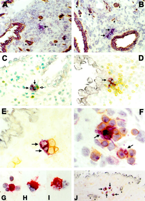
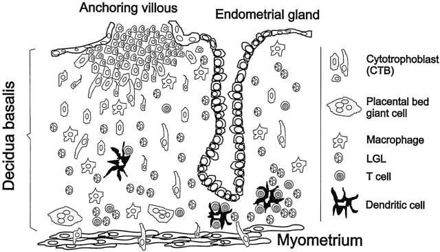
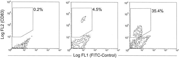
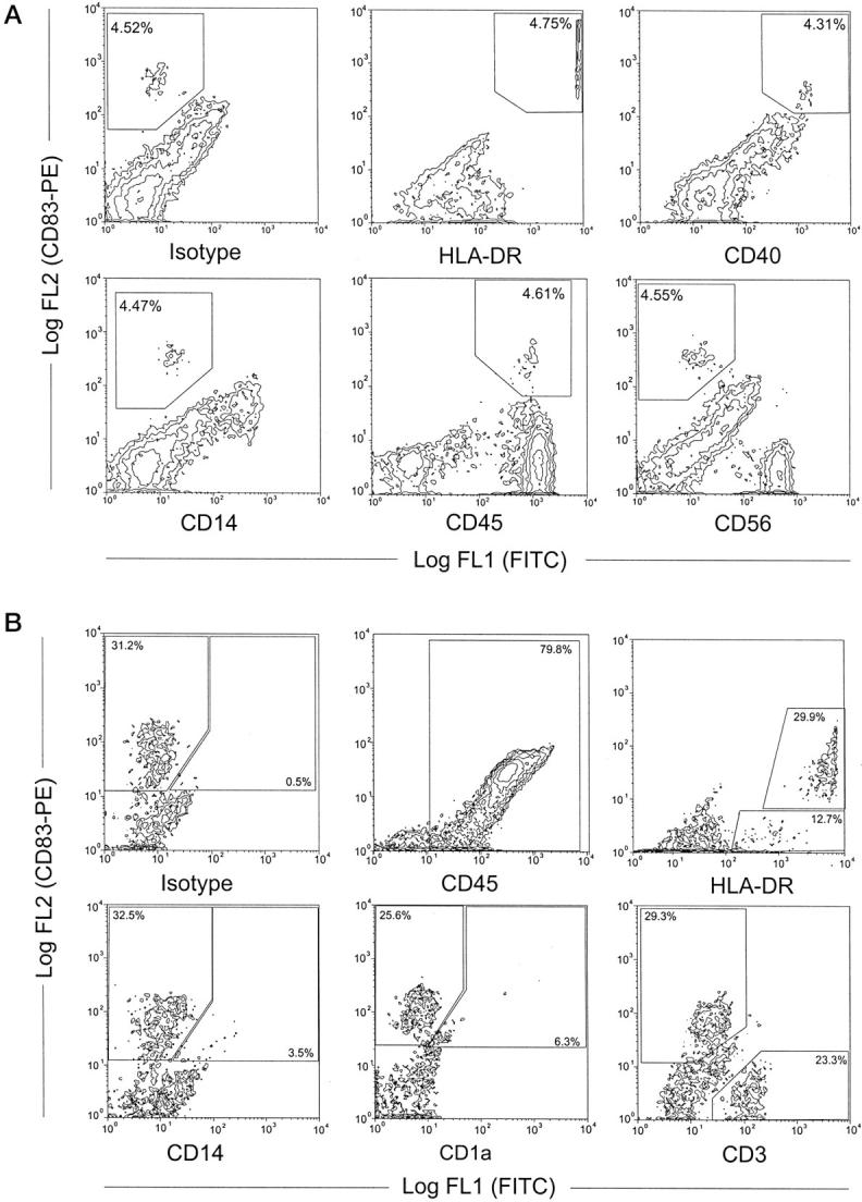
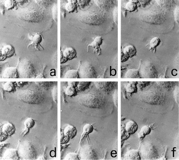
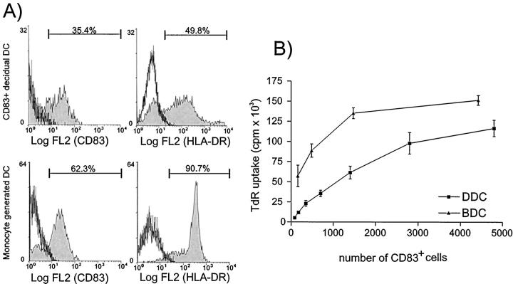
Dendritic cells (DCs) are sentinel cells of the immune system important in initiating antigen-specific T-cell responses to microbial and transplantation antigens. DCs are particularly found in surface tissues such as skin and mucosa, where the organism is threatened by infectious agents. The human decidua, despite its proposed immunosuppressive function, hosts a variety of immunocompetent CD45 cells such as natural killer cells, macrophages, and T cells. Here we describe the detection, isolation, and characterization of CD45(+), CD40(+), HLA-DR(++), and CD83(+) cells from human early pregnancy decidua with typical DC morphology. CD83(+) as well as CD1a(+) cells were found in close vicinity to endometrial glands, with preference to the basal layer of the decidua. In vitro, decidual CD83(+) cells could be enriched to approximately 30%, with the remainder of cells encompassing DC-bound CD3(+) T cells. Stimulation of allogeneic T cells in a mixed leukocyte reaction by the decidual cell fraction enriched for CD83(+) cells, was similar to that obtained with blood monocyte-derived DCs, demonstrating the potent immunostimulatory capacity of these cells. Decidual DCs with morphological, phenotypic, and functional characteristics of immunostimulatory DCs might be important mediators in the regulation of immunological balance between maternal and fetal tissue, leading to successful pregnancy.


Figures

Figure 1.
Figure 1.
Immunohistochemistry of CD83+ and CD1a+ cells in early human pregnancy. Violet; VIP, brown; DAB, gray-blue; Vector blue, red; New fuchsin. A, C, and E: CD1a+ cells (violet, arrows); B, D, F, and J: CD83+ cells (violet, arrows). A: CD1a+ cells in decidua basalis exhibit a typical veiled appearance of DCs with long dendrites extending into the surrounding tissue. Brown: cytokeratin staining of invading FTBs (*) and endometrial glands (original magnification, ×160). B: Most CD83+ cells are located close to cytokeratin-positive endometrial glands (brown; original magnification, ×160). C: Typical location of CD1a+ cells in close vicinity to endometrial glands (gray-blue). CD1a+ cells are associated with a single CD3+ cell (brown, open head arrow; original magnification, ×250). D: CD83+ cells are often associated with CD3+ cells (brown) in clusters near an endometrial gland (gray-blue; original magnification, ×250). E: Association of CD1a+ cells with CD3+ cells (brown; original magnification, ×400). F: Association of CD83+ cells with CD3+ cells (brown; original magnification, ×630). J: CD83+ cells in a lymphatic vessel together with CD45+ lymphocytes (brown). * indicates invading FTBs indicating the tissue as decidua basalis (blue; original magnification, ×63). Cytocentrifuge preparations of enriched CD83+ cells demonstrate them to be often associated with lymphocytes (G; original magnification, ×630), to exhibit typical veiled morphology (H; ×1000) and to express HLA-DR (I; original magnification, ×1000).
Figure 2.
Figure 2.
Scheme of the implantation site. The placental anchoring villous is at the top of the diagram with the FTBs invading the decidua basalis beneath the anchoring villous. Most DCs are located in the basal layer of decidua (as characterized by the presence of the placental bed giant cells) directly at the myometrium at the bottom of the scheme. Most of the DCs are associated with T cells and some LGL.
Figure 3.
Figure 3.
FACS analysis of the enrichment procedure of CD83+ cells from human decidua. Left: decidual mononuclear cells contain 0.2% CD83+ cells. Middle: after overnight culture of the decidual mononuclear cells, the nonadherent fraction contains 4.5% of CD83+ cells. Right: after positive selection, the CD83+ cells are enriched to 35%. Win-MDI-Program, threshold: 0.5, smoothing: 1.
Figure 4.
Figure 4.
A: Double-staining FACS analysis of the phenotype of decidual cells (nonadherent fraction after overnight culture). Approximately 4.5% of the cells are positive for CD83 (upper left) and these co-stain with HLA-DR, CD40, CD45 and, at a very low level, with CD14, but not with CD56. A population of CD83 cells weakly positive for HLA-DR, CD40, and CD14 may represent macrophages, the CD56+ cells (lower right) represent the LGLs typical for human decidua. B: Flow cytometry of immunomagnetically-enriched decidual DC demonstrated 79.8% of the cells to be CD45+, 32.5% to be CD83+, and all CD83+ cells to be highly positive for HLA-DR. A small population of CD83+ cells co-stains with CD14 (3.5%) or CD1a (6.3%). A population of CD83 but weak HLA-DR+ cells (12.7%) may represent immature DC. The main population of the CD83 cells are CD3+ T cells (23.3% of all cells).
Figure 5.
Figure 5.
Morphology and motility of DCs isolated from human decidua over plastic. Monitoring over a time course of one picture per 30 seconds (a–f) demonstrates the motility of cells with characteristic extending and retracting dendrites from the surface of the cells (interference contrast, ×640).
Figure 6.
Figure 6.
A: Phenotype of cells used for the mixed lymphocyte reaction. Top: CD83-enriched cells from human decidua. Bottom: DCs generated from blood monocytes. Cells were labeled as indicated and the percent of positive staining cells is shown. Gates were set using the isotype-matched negative controls to be <0.2% positive. B: Mixed lymphocyte reaction (MLR) of DCs. Graded numbers of CD83+ cells were used as stimulator cells in an allogeneic MLR together with 1 × 10 purified T-lymphocyte responders. Results are shown as the mean [3H]-thymidine uptake (TdR) ± SEM from all six CD83+ decidual DC samples (DDC; squares) and the five blood monocyte-derived DC samples (BDC; triangles). [3H]-thymidine uptake of all decidual DC and BDC populations as well as the responder T cells alone was <500 cpm and is not shown.

References

    1. Billingham RE: Transplantation immunity and the maternal fetal relation. New Engl J Med 1964, 270:667-672 - PubMed
    1. Starkey PM, Sargent IL, Redman CWG: Cell populations in human early pregnancy decidua: characterization and isolation of large granular lymphocytes by flow cytometry. Immunology 1988, 65:129-134 - PMC - PubMed
    1. Bulmer JN, Sunderland CA: Immunohistological characterization of lymphoid cell populations in the early human placental bed. Immunology 1984, 52:349-357 - PMC - PubMed
    1. Mincheva-Nilsson L, Baranov V, Yeung MM, Hammarstrom S, Hammarstrom ML: Immunomorphologic studies of human decidua-associated lymphoid cells in normal early pregnancy. J Immunol 1994, 152:2020-2032 - PubMed
    1. King A, Wellings V, Gardner L, Loke YW: Immunocytochemical characterization of the unusual large granular lymphocytes in human endometrium throughout the menstrual cycle. Hum Immunol 1989, 24:195-205 - PubMed

Publication types