

Antineutrophil cytoplasmic antibodies induce reactive oxygen-dependent dysregulation of primed neutrophil apoptosis and clearance by macrophages
- PMID: 10880391
- PMCID: PMC1850196
- DOI: 10.1016/S0002-9440(10)64532-4
Antineutrophil cytoplasmic antibodies induce reactive oxygen-dependent dysregulation of primed neutrophil apoptosis and clearance by macrophages
Abstract
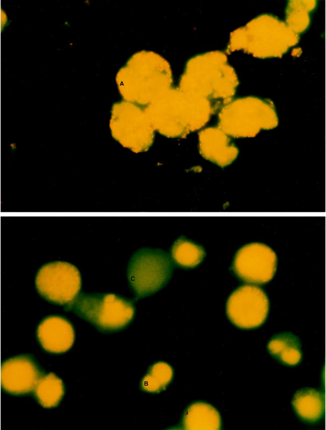
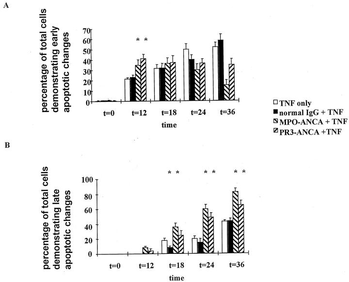
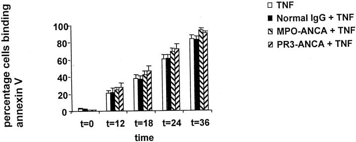
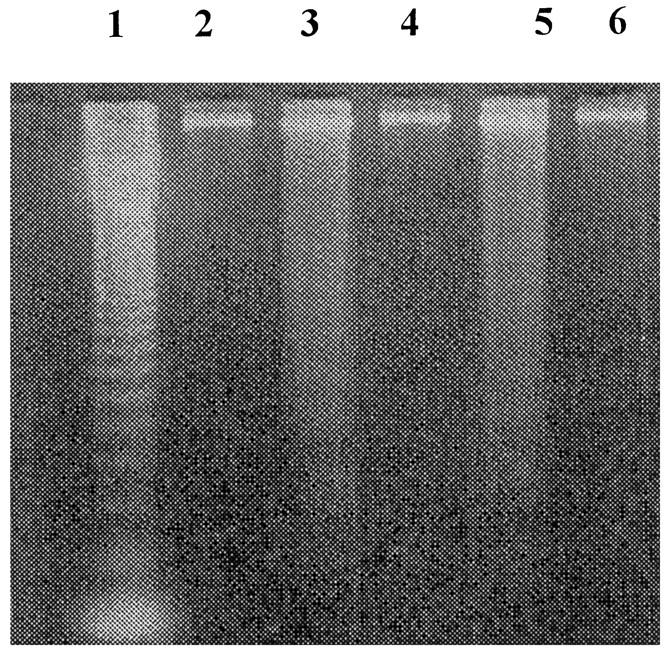
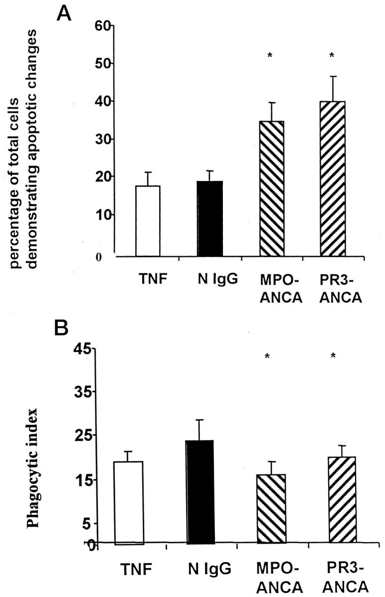
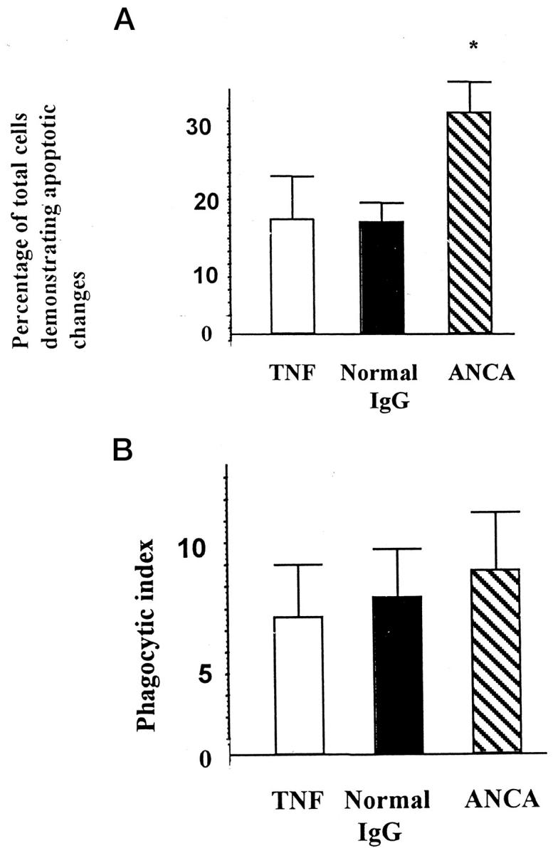
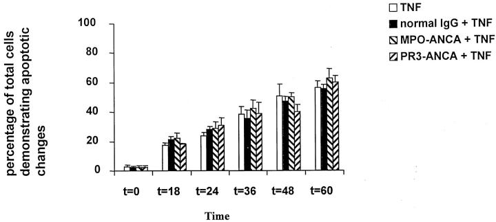
This study assessed whether anti-neutrophil cytoplasmic antibodies (ANCAs) interfere with the safe deletion of neutrophils by apoptosis and phagocytic clearance. Tumor necrosis factor (TNF)-primed neutrophils were incubated with normal IgG (N IgG) or ANCA IgG for up to 36 hours. Compared with N IgG, ANCAs accelerated constitutive apoptosis of TNF-alpha primed neutrophils, as assessed by morphology and confirmed by DNA laddering pattern on gel electrophoresis, and accelerated progression to secondary necrosis. The accelerated apoptosis induced by ANCA was dependent on reactive oxygen species generation, as primed neutrophils from patients with chronic granulomatous disease failed to show an effect of ANCAs on apoptosis. However, there was no change in the rate at which neutrophils exhibited annexin V binding, indicating that externalization of phosphatidylserine was not accelerated by ANCAs. Furthermore, when ANCA-treated primed neutrophils were interacted with human or murine peritoneal macrophages after 12 hours there was significantly less phagocytosis by human macrophages and no difference in phagocytosis by murine peritoneal-derived macrophages when compared with N IgG-treated controls. In conclusion, ANCAs accelerate apoptosis and secondary necrosis in TNF-primed neutrophils by a mechanism dependent on the generation of reactive oxygen species, with uncoupling of nuclear and surface membrane changes, resulting in a "reduced window of opportunity" for phagocytic recognition and engulfment before disintegration.
Figures

Similar articles
-
Anti-myeloperoxidase antibodies enhance phagocytosis, IL-8 production, and glucose uptake of polymorphonuclear neutrophils rather than anti-proteinase 3 antibodies leading to activation-induced cell death of the neutrophils.Clin Rheumatol. 2007 Feb;26(2):216-24. doi: 10.1007/s10067-006-0285-3. Epub 2006 Mar 31. Clin Rheumatol. 2007. PMID: 16575489
-
Neutrophil priming and apoptosis in anti-neutrophil cytoplasmic autoantibody-associated vasculitis.Kidney Int. 2001 May;59(5):1729-38. doi: 10.1046/j.1523-1755.2001.0590051729.x. Kidney Int. 2001. PMID: 11318943
-
Antineutrophil cytoplasm antibody-induced neutrophil nitric oxide production is nitric oxide synthase independent.Kidney Int. 2001 Feb;59(2):593-600. doi: 10.1046/j.1523-1755.2001.059002593.x. Kidney Int. 2001. PMID: 11168940
-
Activation, apoptosis, and clearance of neutrophils in Wegener's granulomatosis.Ann N Y Acad Sci. 2005 Jun;1051:1-11. doi: 10.1196/annals.1361.041. Ann N Y Acad Sci. 2005. PMID: 16126939 Review.
-
Pathogenicity of Proteinase 3-Anti-Neutrophil Cytoplasmic Antibody in Granulomatosis With Polyangiitis: Implications as Biomarker and Future Therapies.Front Immunol. 2021 Feb 18;12:571933. doi: 10.3389/fimmu.2021.571933. eCollection 2021. Front Immunol. 2021. PMID: 33679731 Free PMC article. Review.
Cited by
-
Neutrophil extracellular chromatin traps connect innate immune response to autoimmunity.Semin Immunopathol. 2013 Jul;35(4):465-80. doi: 10.1007/s00281-013-0376-6. Epub 2013 Apr 18. Semin Immunopathol. 2013. PMID: 23595413 Review.
-
Anti-myeloperoxidase antibodies enhance phagocytosis, IL-8 production, and glucose uptake of polymorphonuclear neutrophils rather than anti-proteinase 3 antibodies leading to activation-induced cell death of the neutrophils.Clin Rheumatol. 2007 Feb;26(2):216-24. doi: 10.1007/s10067-006-0285-3. Epub 2006 Mar 31. Clin Rheumatol. 2007. PMID: 16575489
-
Immunopathological aspects of systemic vasculitis.Springer Semin Immunopathol. 2001;23(3):253-65. doi: 10.1007/s002810100074. Springer Semin Immunopathol. 2001. PMID: 11591101 Review. No abstract available.
-
Preventive and therapeutic effects of MG132 by activating Nrf2-ARE signaling pathway on oxidative stress-induced cardiovascular and renal injury.Oxid Med Cell Longev. 2013;2013:306073. doi: 10.1155/2013/306073. Epub 2013 Mar 7. Oxid Med Cell Longev. 2013. PMID: 23533688 Free PMC article. Review.
-
Pathogenesis of ANCA-associated vasculitis.Curr Rheumatol Rep. 2012 Dec;14(6):481-93. doi: 10.1007/s11926-012-0286-y. Curr Rheumatol Rep. 2012. PMID: 22927039 Review.
References
-
- Wyllie A: Glucocorticoid-induced thymocyte apoptosis associated with endogenous endonuclease activation. Nature 1980, 284:555-556 - PubMed
-
- Whyte M, Meagher L, Macdermott J, Haslett C: Impairment of function in ageing neutrophils is associated with apoptosis. J Immunol 1993, 150:5124-5134 - PubMed
-
- Cox G, Crossley J, Xing Z: Macrophage engulfment of apoptotic neutrophils contributes to the resolution of acute pulmonary inflammation in vivo. Am J Respir Cell Mol Biol 1995, 12:232–237 - PubMed
MeSH terms
Substances
LinkOut - more resources
Full Text Sources
