

Multiple sclerosis and chronic autoimmune encephalomyelitis: a comparative quantitative study of axonal injury in active, inactive, and remyelinated lesions
- PMID: 10880396
- PMCID: PMC1850217
- DOI: 10.1016/S0002-9440(10)64537-3
Multiple sclerosis and chronic autoimmune encephalomyelitis: a comparative quantitative study of axonal injury in active, inactive, and remyelinated lesions
Abstract
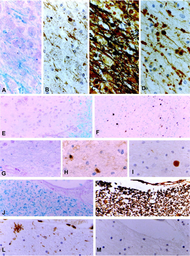
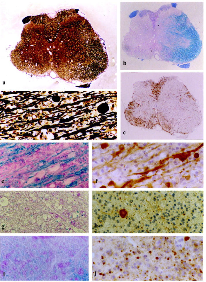
Recent magnetic resonance (MR) studies of multiple sclerosis lesions indicate that axonal injury is a major correlate of permanent clinical deficit. In the present study we systematically quantified acute axonal injury, defined by immunoreactivity for beta-amyloid-precursor-protein in dystrophic neurites, in the central nervous system of 22 multiple sclerosis patients and 18 rats with myelin-oligodendrocyte glycoprotein (MOG)-induced chronic autoimmune encephalomyelitis (EAE). The highest incidence of acute axonal injury was found during active demyelination, which was associated with axonal damage in periplaque and in the normal appearing white matter of actively demyelinating cases. In addition, low but significant axonal injury was also observed in inactive demyelinated plaques. In contrast, no significant axonal damage was found in remyelinated shadow plaques. The patterns of axonal pathology in chronic active EAE were qualitatively and quantitatively similar to those found in multiple sclerosis. Our studies confirm previous observations of axonal destruction in multiple sclerosis lesions during active demyelination, but also indicate that ongoing axonal damage in inactive lesions may significantly contribute to the clinical progression of the disease. The results further emphasize that MOG-induced EAE may serve as a suitable model for testing axon-protective therapies in inflammatory demyelinating conditions.
Figures
References
-
- Charcot JM: Histologie de la sclérose en plaques. Gazette des Hopitaux civils et militaires 1868, 140:554–555 and 141:557–558 and 143:566
-
- Arnold DL, Riess GT, Matthews PM, Francis GS, Collins DL, Wolfson C, Antel JP: Use of proton magnetic resonance spectroscopy for monitoring disease progression in multiple sclerosis. Ann Neurol 1994, 36:76-82 - PubMed
-
- Barnes D, Munro PMG, Youl BD: The longstanding MS lesion: a quantitative MRI and electron microscopic study. Brain 1991, 114:1271-1280 - PubMed
-
- Davie CA, Hawkins CP, Barker GJ, Brennan A, Tofts PS, Miller DH, McDonald WI: Serial proton magnetic resonance spectroscopy in acute multiple sclerosis lesions. Brain 1994, 117:49-58 - PubMed
-
- Davie CA, Barker GJ, Webb S, Tofts PS, Thompson AJ, Harding AE, McDonald WI, Miller DH: Persistent functional deficit in multiple sclerosis and autosomal dominant cerebellar ataxia is associated with axonal loss. Brain 1995, 118:1583-1592 - PubMed
Publication types
MeSH terms
Substances
LinkOut - more resources
Full Text Sources
Other Literature Sources
Medical
