. 2000 Jul;157(1):277-86.
doi: 10.1016/s0002-9440(10)64538-5.

Endocytic pathway abnormalities precede amyloid beta deposition in sporadic Alzheimer's disease and Down syndrome: differential effects of APOE genotype and presenilin mutations

Affiliations

Endocytic pathway abnormalities precede amyloid beta deposition in sporadic Alzheimer's disease and Down syndrome: differential effects of APOE genotype and presenilin mutations

A M Cataldo et al. Am J Pathol. 2000 Jul.

Abstract

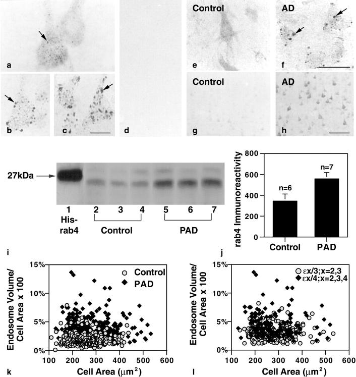
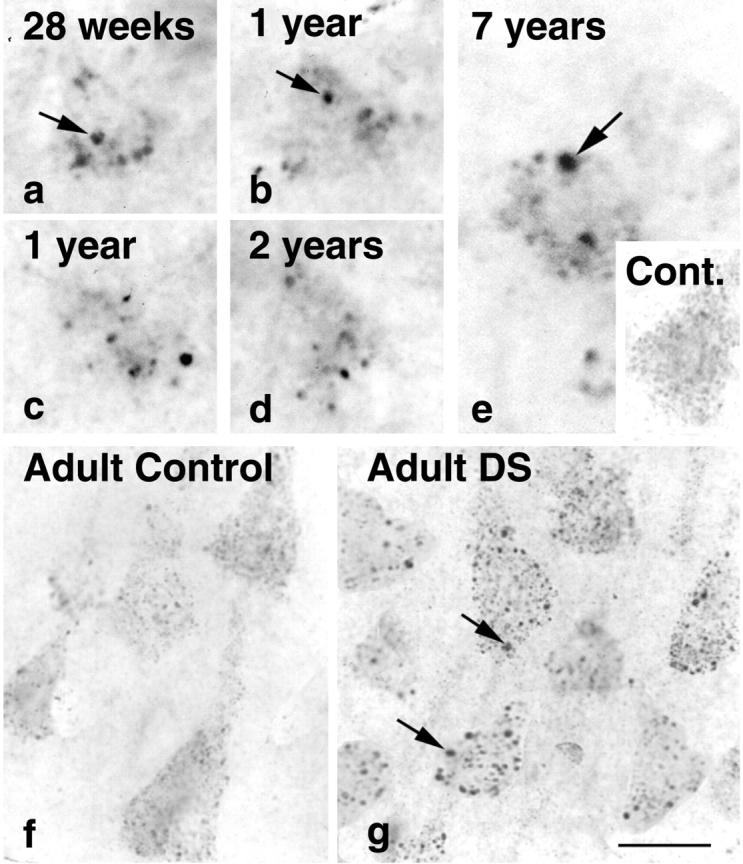
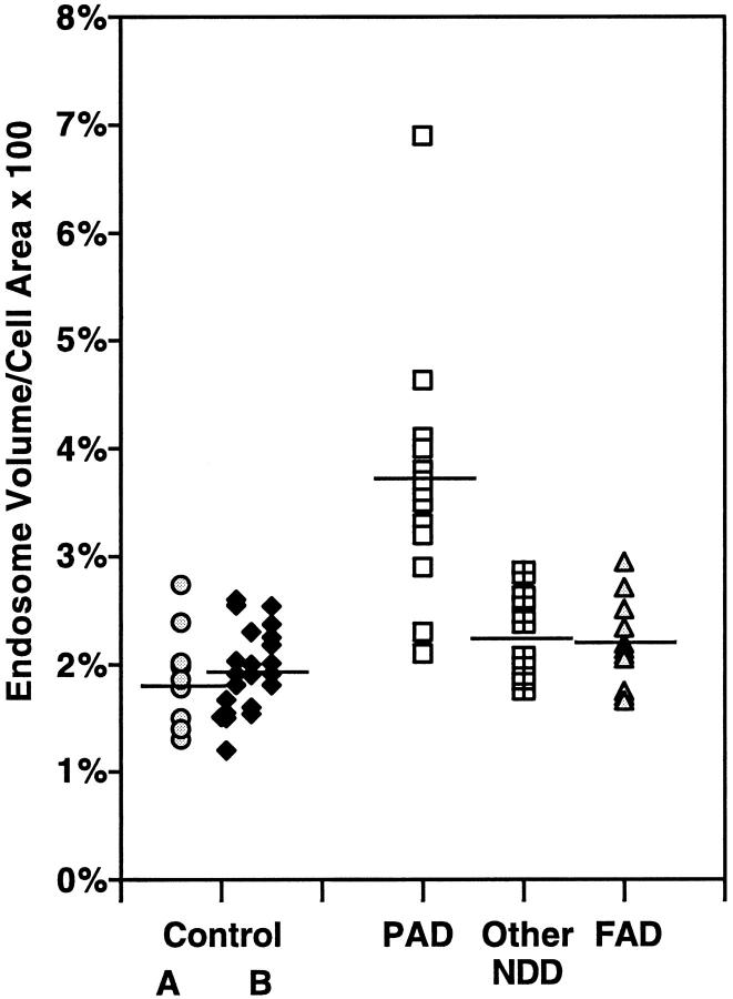
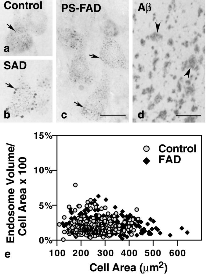
Endocytosis is critical to the function and fate of molecules important to Alzheimer's disease (AD) etiology, including the beta protein precursor (betaPP), amyloid beta (Abeta) peptide, and apolipoprotein E (ApoE). Early endosomes, a major site of Abeta peptide generation, are markedly enlarged within neurons in the Alzheimer brain, suggesting altered endocytic pathway (EP) activity. Here, we show that neuronal EP activation is a specific and very early response in AD. To evaluate endocytic activation, we used markers of internalization (rab5, rabaptin 5) and recycling (rab4), and found that enlargement of rab5-positive early endosomes in the AD brain was associated with elevated levels of rab4 immunoreactive protein and translocation of rabaptin 5 to endosomes, implying that both endocytic uptake and recycling are activated. These abnormalities were evident in pyramidal neurons of the neocortex at preclinical stages of disease when Alzheimer-like neuropathology, such as Abeta deposition, was restricted to the entorhinal region. In Down syndrome, early endosomes were significantly enlarged in some pyramidal neurons as early as 28 weeks of gestation, decades before classical AD neuropathology develops. Markers of EP activity were only minimally influenced by normal aging and other neurodegenerative diseases studied. Inheritance of the epsilon4 allele of APOE, however, accentuated early endosome enlargement at preclinical stages of AD. By contrast, endosomes were normal in size at advanced stages of familial AD caused by mutations of presenilin 1 or 2, indicating that altered endocytosis is not a consequence of Abeta deposition. These results identify EP activation as the earliest known intraneuronal change to occur in sporadic AD, the most common form of AD. Given the important role of the EP in Abeta peptide generation and ApoE function, early endosomal abnormalities provide a mechanistic link between EP alterations, genetic susceptibility factors, and Abeta generation and suggest differences that may be involved in Abeta generation and beta amyloidogenesis in subtypes of AD.


Figures

Figure 1.
Figure 1.
Early endosomal alterations are the earliest detectable intracellular manifestation of AD and are evident in individuals who have not yet developed clinical symptoms. Pyramidal neurons of the prefrontal cortex of an early-stage sporadic AD case labeled with rab5 (b) and EEA1 (c) show atypically large endosomes (arrows). a: rab5-immunopositive neuronal early endosomes (arrow) from neurons in an age-matched control. Serial adjacent sections immunolabeled with an antibody to Aβ17–24 (d) or MC-1 (not shown) displayed minimal to no evidence of extracellular Aβ deposition or neurofibrillary pathology. Immunolabeling for rabaptin 5, a regulator protein of endocytosis, was detected in the cytosol and associated with small vesicles in control brains (e). By contrast, rabaptin5 immunoreactivity in neurons from pre-AD brains was predominantly located on large endosomes (f, arrows), a change indicative of increased endosomal fusion. Compared with neurons from control brains (g), pyramidal neurons in the pre-AD cortex (h) showed increased levels of rab4 immunoreactivity, which is consistent with an increase in endosome to plasma membrane vesicular recycling. Western blot analysis (i) of brain homogenates (100 μg/lane) prepared from the frontal cortex of three representative neuropathologically-normal controls (n = 6; lanes 2–4) and three representative pre-AD brains (n = 7) (PAD, lanes 5–7) confirmed the immunocytochemical findings and revealed an increase in rab4 immunoreactive protein (Mr ∼23 to 25) in the pre-AD brains. (Lane 1, recombinant His-tagged rab4; CytoSignal Research Products, Irvine CA.) Scan analysis (j) of the rab4 protein levels obtained by Western blot showed approximately twofold higher rab4 levels in pre-AD brains compared with control cases (control mean = 345.1; pre-AD mean = 560.6; P > 0.05. Error bars represent SEM. Morphometric analysis (k) of 25 pyramidal neurons from each of 10 control and 15 early-stage AD brains showed an average twofold larger total endosomal volume per neuron in the AD cases versus control, implying an increase in endocytosis (possible AD mean = 3.68%, SEM ± 0.093; control mean = 1.88%, SEM ± 0.066; P < 0.005). Reassessment (l) of the same early SAD cases, stratified by APOE genotype, showed approximately twofold higher endosomal volume per neuron in lamina III pyramids of the five brains carrying one or both copies of the ε4 allele of APOE versus the 10 brains with ε2 or ε3 alleles (ε4 mean = 4.68, SEM ± 0.206;ε2, ε3 mean = 3.18, ± SEM 0.095, P < 0.005). Scale bars: a–c, 20 μm; e and f, 50 μm; g and h, 200 μm.
Figure 2.
Figure 2.
Early endosomal enlargement is evident in single neurons from the prefrontal cortices of a fetus (a), infants of 1 and 2 years (b–d), and a young 7-year-old child (e) with DS immunolabeled with rab5 (a–e). The numbers of neurons containing enlarged endosomes increased with the age of the individual. The magnitude of endosomal enlargement in neurons detected with rab5 in young DS individuals did not differ significantly from that observed in cases of adult DS (g). e, inset, and f show representative neurons from young and aged control brains, respectively. Scale bars: a–g, 20 μm
Figure 3.
Figure 3.
Morphometric analyses of neuronal endosomal volume in neurodegenerative diseases and normal individuals of varying age. The values are expressed as the percentage of cell area occupied by rab5-positive early endosomes averaged for 25 pyramidal neurons in each brain indicated by a data point on the graph. Results show endosomal enlargement is specific to non-PS forms of AD (elderly, nondemented control group A, mean = 1.88%, SEM ± 0.066; pre-AD/PAD mean = 3.68%, SEM ± 0.093; other non-AD neurodegenerative disorders, mean = 2.26%, SEM ± 0.067; PS-FAD mean = 2.22, SEM ± 0.062). Control group B represents a second pool of 22 neurologically normal individuals of ascending age comprising four groups: 1 to 15 years, n = 5, mean = 1.45%, SEM ± 0.095; 16 to 35 years, n = 5, mean = 2.19%, SEM ± 0.062; 36 to 55 years, n = 5, mean = 1.86%, SEM ± 0.077; and >55 years, n = 7, mean = 2.14%, SEM ± 0.094.
Figure 4.
Figure 4.
Neuronal endosomes in individuals with PS mutations (c) are comparable in size to those in age-matched controls (a) despite high levels of parenchymal Aβ deposition in the PS-FAD brains (d, arrowheads). By contrast, most pyramidal neurons in sporadic Alzheimer brain display large endosomes (b, arrow). e: Morphometric analysis of early endosomal size from each of 10 control and 15 PS-FAD brains. Expressed as percent cell area occupied by rab5-positive early endosomes, the results show no significant difference in endosomal volume in the PS-FAD brains versus control (PS-FAD mean = 2.22%, SEM ± 0.06; control mean = 1.88%, SEM ± 0.07). Scale bars: a–c, 20 μm; d, 200 μm.

References

    1. Chyung ASC, Greenberg BD, Cook DG, Doms RW, Lee VM: Novel beta-secretase cleavage of beta-amyloid precursor protein in the endoplasmic reticulum/intermediate compartment of NT2N cells. J Cell Biol 1997, 138:671-680 - PMC - PubMed
    1. Cook DG, Forman MS, Sung JC, Leight S, Kolson DL, Iwatsubo T, Lee VM, Doms RW: Alzheimer’s A beta(1–42) is generated in the endoplasmic reticulum/intermediate compartment of NT2N cells. Nat Med 1997, 3:1021-1023 - PubMed
    1. Hartmann H, Busciglio J, Baumann KH, Staufenbiel M, Yankner BA: Developmental regulation of presenilin-1 processing in the brain suggests a role in neuronal differentiation. J Biol Chem 1997, 272:14505-14508 - PubMed
    1. Xia W, Zhang J, Ostaszewski BL, Kimberly WT, Seubert P, Koo EH, Shen J, Selkoe DJ: Presenilin 1 regulates the processing of beta-amyloid precursor protein C-terminal fragments and the generation of amyloid beta-protein in endoplasmic reticulum and Golgi. Biochemistry 1998, 37:16465-16471 - PubMed
    1. Xu H, Sweeney D, Wang R, Thinakaran G, Lo AC, Sisodia SS, Greengard P, Gandy S: Generation of Alzheimer beta-amyloid protein in the trans-Golgi network in the apparent absence of vesicle formation. Proc Natl Acad Sci USA 1997, 94:3748-3752 - PMC - PubMed

Publication types

MeSH terms