. 2000 Jul 3;19(13):3168-78.
doi: 10.1093/emboj/19.13.3168.

Crystal structure of cystalysin from Treponema denticola: a pyridoxal 5'-phosphate-dependent protein acting as a haemolytic enzyme

Affiliations

Crystal structure of cystalysin from Treponema denticola: a pyridoxal 5'-phosphate-dependent protein acting as a haemolytic enzyme

H I Krupka et al. EMBO J. .

Abstract

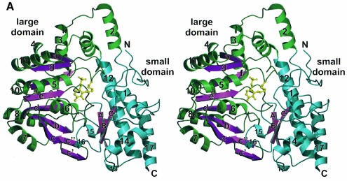
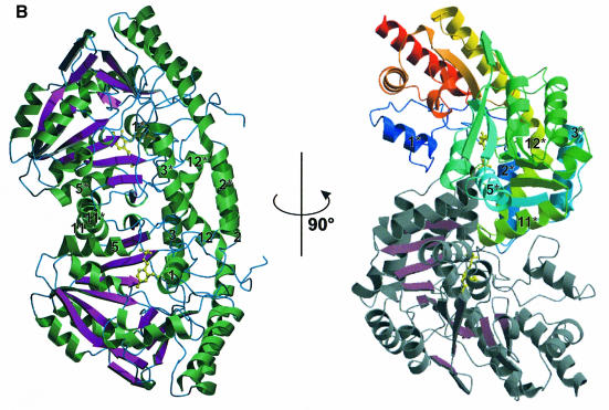
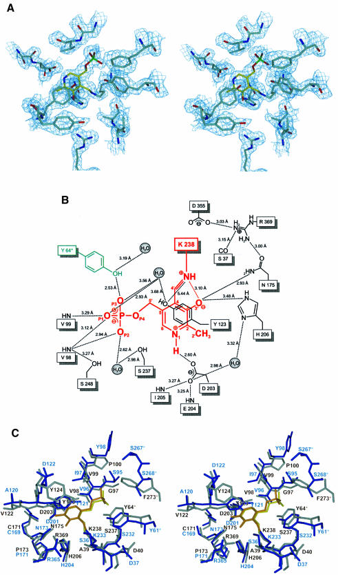
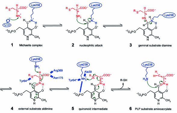
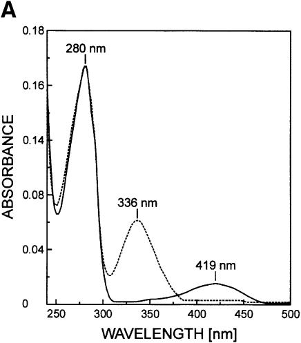
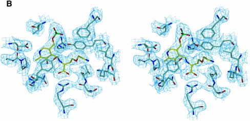
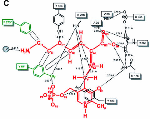
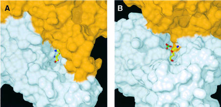
Cystalysin is a C(beta)-S(gamma) lyase from the oral pathogen Treponema denticola catabolyzing L-cysteine to produce pyruvate, ammonia and H(2)S. With its ability to induce cell lysis, cystalysin represents a new class of pyridoxal 5'-phosphate (PLP)-dependent virulence factors. The crystal structure of cystalysin was solved at 1.9 A resolution and revealed a folding and quaternary arrangement similar to aminotransferases. Based on the active site architecture, a detailed catalytic mechanism is proposed for the catabolism of S-containing amino acid substrates yielding H(2)S and cysteine persulfide. Since no homologies were observed with known haemolysins the cytotoxicity of cystalysin is attributed to this chemical reaction. Analysis of the cystalysin-L-aminoethoxyvinylglycine (AVG) complex revealed a 'dead end' ketimine PLP derivative, resulting in a total loss of enzyme activity. Cystalysin represents an essential factor of adult periodontitis, therefore the structure of the cystalysin-AVG complex may provide the chemical basis for rational drug design.


Figures

None
Fig. 1. Overall fold. (A) Structure of the cystalysin monomer showing the large domain with green helices and magenta β-sheets, the small domain with blue helices and the PLP cofactor in yellow. The nomenclature for the secondary structural elements is indicated. (B) Ribbon presentation of the cystalysin dimer emphasizing structural elements essential for dimerization. Helical structures are shown in green, loop structure in blue and β-sheets in magenta. 90° rotation results in the characteristic S-shaped orientation in which the sequential flow is mapped by colour ramp to one monomer (N-terminus, blue; C-terminus, red).
None
Fig. 2. Active site structure. (A) Final 2FoFc electron density of the PLP cofactor (yellow) and the immediate protein vicinity (colour coding by atom type), contoured at 1.2σ and calculated at 1.9 Å resolution. (B) Schematic presentation of the active site around the PLP cofactor (red). Residues contributing from the neighbouring subunit are coloured green. Hydrogen bonds and interatomic distances (Å) are indicated. (C) Stereo plot showing the superposition of the active sites of cystalysin and MalY from E.coli. Cystalysin residues are coloured grey with the cofactor in yellow, MalY is shown in blue and its PLP in orange.
None
Fig. 3. Proposed molecular reaction mechanism for cystalysin showing relevant intermediates. The PLP cofactor is coloured black, the substrate and leaving product in red and the enzyme residues in blue.
None
Fig. 4. The cystalysin–AVG complex. (A) The spectra of uncomplexed cystalysin and cystalysin after treatment with AVG, recorded at 20°C in 100 mM phosphate buffer. (B) Final 2FoFc electron density of the PLP–AVG complex (yellow) and the immediate protein vicinity (colour coding by atom type), contoured at 1.2σ and calculated at 2.5 Å resolution. (C) Schematic presentation of the PLP–AVG complex (red) depicted with polar and hydrophobic interactions. Interatomic distances are given in Å.
None
Fig. 5. Active site morphology. Surface plots of the active site cavities of cystalysin (A) and CBL (B) both with the PLP–AVG complex in yellow (colour coding by atom type; P, green). The surface of the subunit, harbouring the complex, is coloured in grey, the neighbouring subunit in orange.

References

    1. Alexeev D., Alexeeva,M., Baxter,R.L., Campopiano,D., Webster,S.P. and Sawyer,L. (1998) The crystal structure of 8-amino-7-oxononoate synthase: a bacterial PLP-dependent Acyl-CoA-condensing enzyme. J. Mol. Biol., 284, 401–419. - PubMed
    1. Beauchamp R.O., Bus,J.S., Popp,J.A., Boreiko,C.J. and Andjelkovich,D.A. (1984) A critical review of the literature on hydrogen sulfide toxicity. Crit. Rev. Toxicol., 13, 25–97. - PubMed
    1. Blankenfeldt W., Nowicki,C., Hunter,G.R., Montemartini-Kalisz,M., Kalisz,H.M. and Hecht,H.J. (1999) Crystal structure of Trypanosoma cruzi tyrosine aminotransferase: substrate specificity is influenced by cofactor binding mode. Protein Sci., 8, 2406–2417. - PMC - PubMed
    1. Brünger A.T. et al. (1998) Crystallography and NMR system: a new software suite for macromolecular structure determination. Acta Crystallogr. D, 54, 905–921. - PubMed
    1. Chu L. and Holt,S.C. (1994) Purification and characterization of a 45 kDa hemolysin from Treponema denticola ATCC 35404. Microb. Pathog., 16, 197–212. - PubMed