. 2000 Jul 3;19(13):3235-49.
doi: 10.1093/emboj/19.13.3235.

Salmonella maintains the integrity of its intracellular vacuole through the action of SifA

Affiliations

Salmonella maintains the integrity of its intracellular vacuole through the action of SifA

C R Beuzón et al. EMBO J. .

Erratum in

  • EMBO J 2000 Aug 1;19(15):4191

Abstract

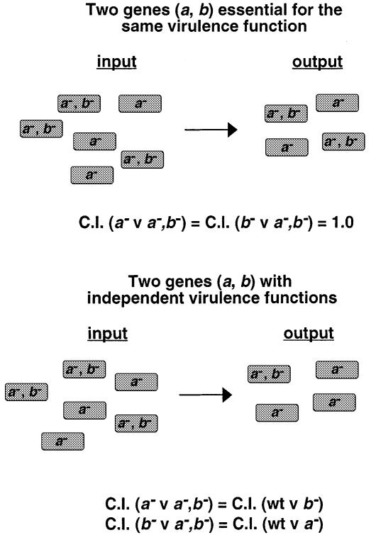
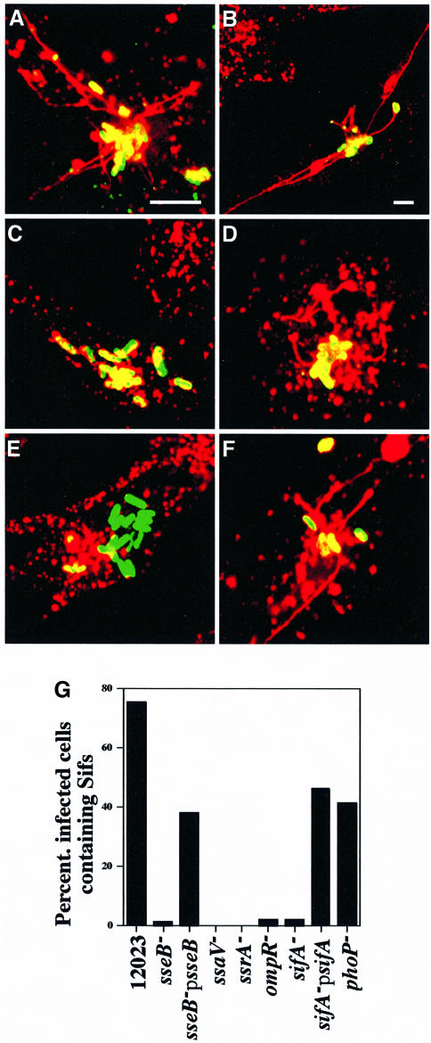
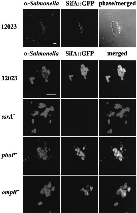
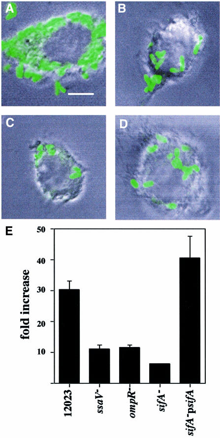
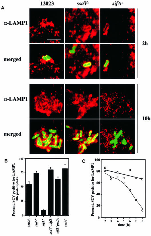
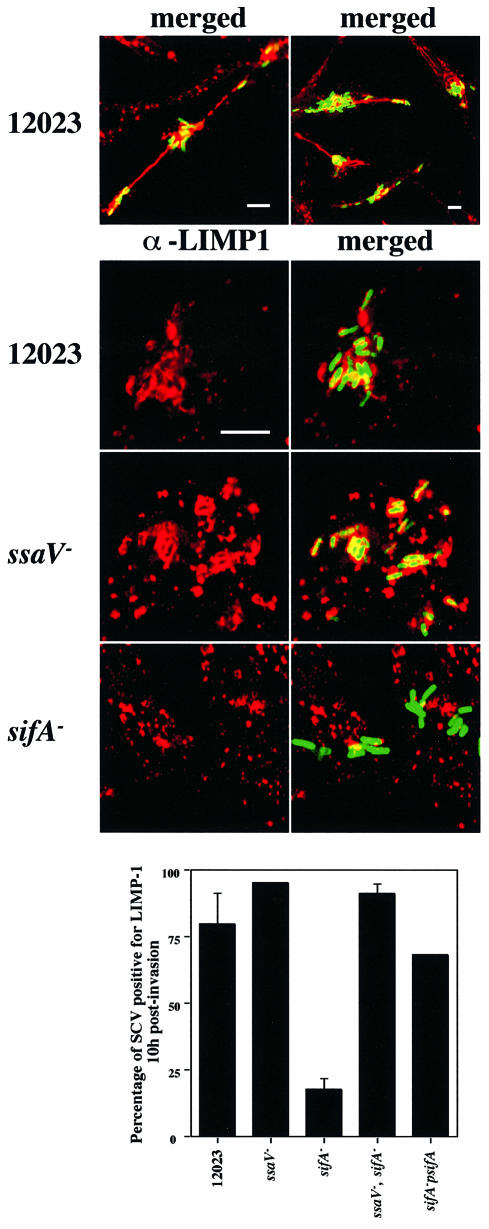
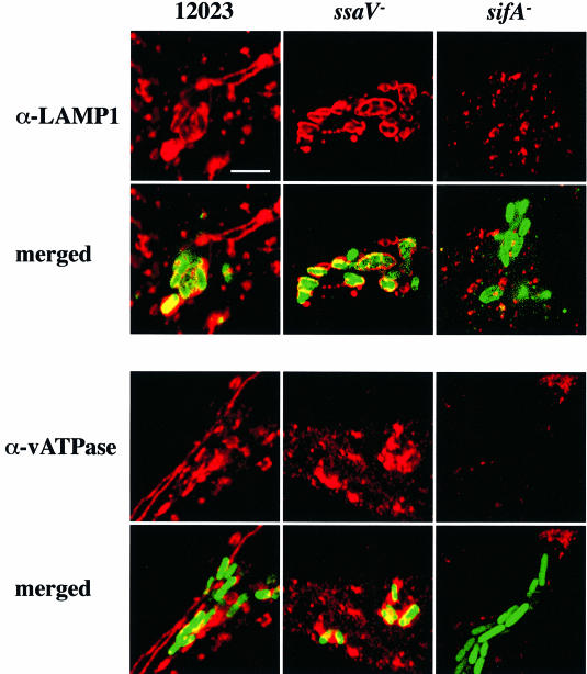
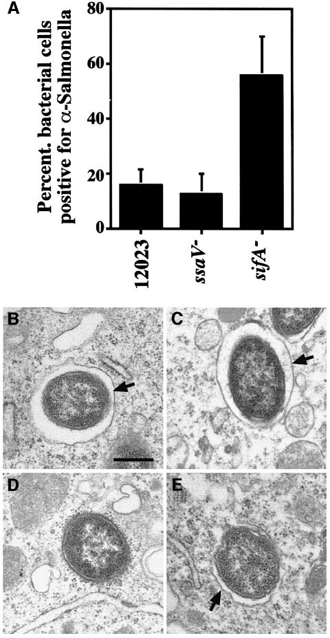
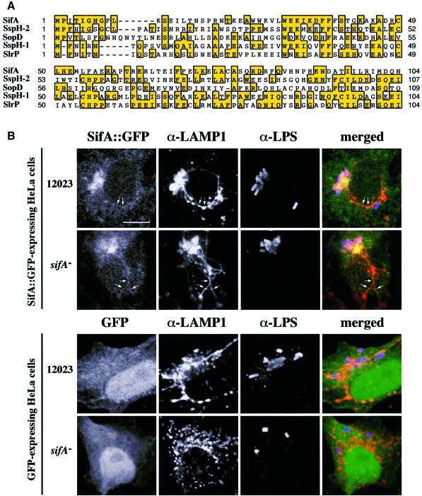
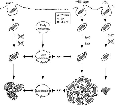
A method based on the Competitive Index was used to identify Salmonella typhimurium virulence gene interactions during systemic infections of mice. Analysis of mixed infections involving single and double mutant strains showed that OmpR, the type III secretion system of Salmonella pathogenicity island 2 (SPI-2) and SifA [required for the formation in epithelial cells of lysosomal glycoprotein (lgp)-containing structures, termed Sifs] are all involved in the same virulence function. sifA gene expression was induced after Salmonella entry into host cells and was dependent on the SPI-2 regulator ssrA. A sifA(-) mutant strain had a replication defect in macrophages, similar to that of SPI-2 and ompR(-) mutant strains. Whereas wild-type and SPI-2 mutant strains reside in vacuoles that progressively acquire lgps and the vacuolar ATPase, the majority of sifA(-) bacteria lost their vacuolar membrane and were released into the host cell cytosol. We propose that the wild-type strain, through the action of SPI-2 effectors (including SpiC), diverts the Salmonella-containing vacuole from the endocytic pathway, and subsequent recruitment and maintenance of vacuolar ATPase/lgp-containing membranes that enclose replicating bacteria is mediated by translocation of SifA.


Figures

None
Fig. 1. Theoretical representation of Competitive Index (C.I.) analysis. C.I. is the c.f.u. ratio of double and single strains recovered from the infected animal divided by the c.f.u. ratio of double and single strains in the inoculum. Panels represent different degrees of functional relationship between two hypothetical genes (a and b) and the C.I. predicted for each case. In this case:
None
Fig. 2. Sif formation by S.typhimurium mutant strains in HeLa cells. Confocal immunofluorescence analysis of Sif formation in cells infected with: 12023 (A and B), sseB (C), sseB psseB (D), sifA (E) and sifApsifA (F) strains. Cells were fixed at 7 h post-infection and stained with mouse anti-LAMP-1 and TRSC-conjugated donkey anti-mouse antibodies. In (A–E) the bacterial strains carried a plasmid expressing gfp constitutively. In (F), bacterial cells were detected by goat anti-Salmonella and FITC-conjugated donkey anti-goat antibodies. sseB and sifA mutant strains fail to induce Sifs (C and E). This defect can be complemented by the introduction of plasmids carrying the respective wild-type alleles (D and F). Bars correspond to 4 µm; the scale in (C–F) is equivalent to that of (A). (G) represents typical results from one of three experiments where cells (n = 50) infected by each S.typhimurium strain were evaluated for Sif formation at 7 h post-invasion. Values are given as percentage of infected cells containing Sifs.
None
Fig. 3. sifA::gfp expression by S.typhimurium mutant strains in infected HeLa cells. Confocal immunofluorescence analysis in cells 6 h after invasion with different strains carrying a plasmid-borne sifA::gfp fusion (pID812). Salmonella typhimurium was detected with goat anti-Salmonella, and TRSC-conjugated donkey anti-goat antibodies. The upper panel shows three extracellular bacteria not expressing GFP, and several GFP-positive intracellular bacteria. SifA is expressed within HeLa cells in an ssrA-dependent manner (middle panel). SifA expression is not affected by a phoP mutation but was only detected in ∼50% of bacterial cells carrying the ompR mutation. Bars correspond to 4 µm.
None
Fig. 4. A sifA mutant strain is defective for replication within RAW macrophages. Opsonized bacteria were taken up by phagocytosis and at 2 and 16 h host cells were either lysed and cultured for enumeration of intracellular bacteria (gentamicin-protected), or fixed and examined by phase-contrast and confocal fluorescence microscopy. (A, B, C and D) represent typical cells infected with GFP-expressing 12023, ssaV, sifA and ompR, respectively. In (E), the values shown represent the fold increase calculated as a ratio of the intracellular bacteria between 2 and 16 h post-uptake. Each strain was infected in triplicate and the standard errors from the means are shown. The results shown are representative of three independent experiments. Scale bar represents 4 µm.
None
Fig. 5. The lysosomal membrane protein LAMP-1 is not associated with sifA mutant bacteria 10 h post-uptake in RAW macrophages. Confocal microscopic analysis was carried out on macrophages infected with wild-type (12023), ssaV or sifA strains constitutively expressing GFP, at 2 h [upper panels (A)] or 10 h [lower panels (A)] post-uptake. LAMP-1 was detected using a rabbit polyclonal and TRSC-conjugated donkey anti-rabbit secondary antibodies. Scale bar corresponds to 4 µm. The percentage of bacteria (n = 50) co-localizing with LAMP-1 was determined for these and two other bacterial strains 10 h after uptake. The percentage of bacteria (n = 50) co-localizing with LAMP-1 at different time points was determined for 12023 (open squares) and sifA mutant (open circles) strains (C). Results shown in (B) are the means ± SE of three independent experiments. For the sifA, psifA strain, bacteria were detected with goat anti-Salmonella and FITC-conjugated donkey anti-goat antibodies.
None
Fig. 6. LIMP-1 is a component of Sifs and is not associated with sifA mutant bacteria 10 h after invasion of HeLa cells. Confocal microscopic analysis was carried out on cells infected with wild-type (12023), ssaV or sifA strains constitutively expressing GFP. LIMP-1 was detected using a mouse mAb and TRSC-conjugated donkey anti-mouse secondary antibodies. Low magnification images show that LIMP-1 is a component of Sifs in cells infected with wild-type bacteria (upper panel). Higher magnification images show LIMP-1-containing SCVs for wild-type and ssaV mutant bacteria, but weak or no association of LIMP-1 with sifA mutant cells. Scale bars correspond to 4 µm. The percentage of bacteria (n = 50) co-localizing with LIMP-1 was determined for these and two other bacterial strains. Results shown are the means ± SE of three independent experiments. For the sifA, psifA strain, bacteria were detected with goat anti-Salmonella followed by FITC-conjugated donkey anti-goat antibodies.
None
Fig. 7. LAMP-1 and vATPase are not associated with sifA mutant bacteria 10 h post-invasion in HeLa cells. Confocal microscopic analysis was carried out on cells infected with wild-type (12023), ssaV or sifA strains constitutively expressing GFP. LAMP-1 and vATPase were detected using mouse monoclonal antibodies, followed by TRSC-conjugated donkey anti-mouse secondary antibodies. Scale bar corresponds to 4 µm.
None
Fig. 8. Streptolysin-O permeabilization and electron microscopy of host cells infected with different bacterial strains. (A) RAW cells were infected with different strains expressing GFP for 8 h, then permeabilized with streptolysin-O, and incubated with anti-LPS antibody. The percentage of bacteria staining positive for the anti-LPS antibody was determined. Results shown are the means ± SE of three independent experiments. In each experiment, between 100 and 150 bacteria were scored for each strain. Electron microscopy analysis of RAW macrophages infected with either wild-type (B), ssaV (C), sifA (D) or sifA, psifA (E) strains, 10 h post-uptake. Infected cells were fixed and processed for transmission electron microscopy. Arrows in (B, C and E) point to vacuolar membranes, which are absent in (D). Scale bar corresponds to 0.5 µm.
None
Fig. 9. (A) Multiple alignment of the N-terminal region of SifA with other Salmonella proteins. The alignment was generated by ClustalW (http://www2.ebi.ac.uk/clustalw/) and Multalin (http://www.toulouse.inra.fr/multalin.html) and edited by hand. (B) Formation of Sifs is restored in sifA mutant-infected cells expressing SifA::GFP. HeLa cells transiently expressing SifA::GFP (upper panels) or GFP as a control (lower panels) were infected with wild-type (12023) or sifA strains, and fixed with paraformaldehyde at 8 h post-infection. LAMP-1 and LPS were detected using TRSC- and Cy5-conjugated donkey anti-rabbit and anti-mouse secondary antibodies, respectively. Three-colour confocal microscopic analysis was performed. Scale bar represents 10 µm. Expression of SifA::GFP in HeLa cells restores the presence of a LAMP-1-enriched vacuole around sifA bacteria. SifA–GFP is localized on SCVs and Sifs (arrows in the top panels).
None
Fig. 10. Model showing the role of the SPI-2 secretion system in the maturation of the SCV.

References

    1. Altschul S., Madden,T., Schaffer,A., Zhang,J., Zhang,Z., Miller,W. and Lipman,D. (1997) Gapped blast and PSI-blast: a new generation of protein database search programs. Nucleic Acids Res., 25, 3389–3402. - PMC - PubMed
    1. Baumler A., Tsolis,R., Valentine,P., Ficht,T. and Heffron,F. (1997) Synergistic effect of mutations in invA and lpfC on the ability of Salmonella typhimurium to cause murine typhoid. Infect. Immun., 65, 2254–2259. - PMC - PubMed
    1. Beuzón C., Banks,G., Deiwick,J., Hensel,M. and Holden,D.W. (1999) pH-dependent secretion of SseB, a product of the SPI-2 type III secretion system of Salmonella typhimurium. Mol. Microbiol., 33, 806–816. - PubMed
    1. Blanc-Potard A. and Groisman,E. (1997) The Salmonella selC locus contains a pathogenicity island mediating intramacrophage survival. EMBO J., 16, 5376–5385. - PMC - PubMed
    1. Chang A.C. and Cohen,S.N. (1978) Construction and characterisation of amplifiable multicopy DNA cloning vehicles derived from the P15A cryptic miniplasmid. J. Bacteriol., 134, 1141–1156. - PMC - PubMed

Publication types

MeSH terms