. 2000 Jul 3;19(13):3272-82.
doi: 10.1093/emboj/19.13.3272.

Thrombospondin-1 suppresses wound healing and granulation tissue formation in the skin of transgenic mice

Affiliations

Thrombospondin-1 suppresses wound healing and granulation tissue formation in the skin of transgenic mice

M Streit et al. EMBO J. .

Abstract

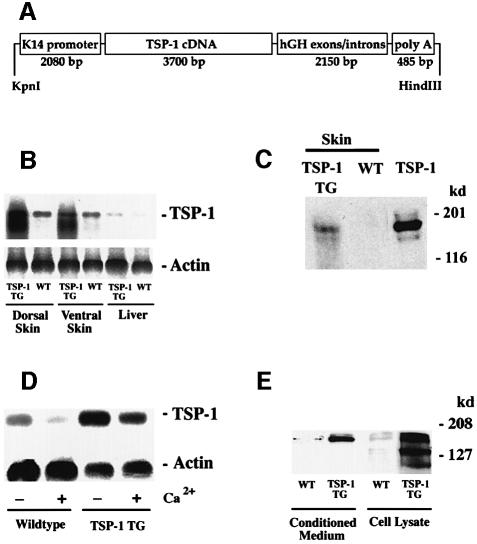
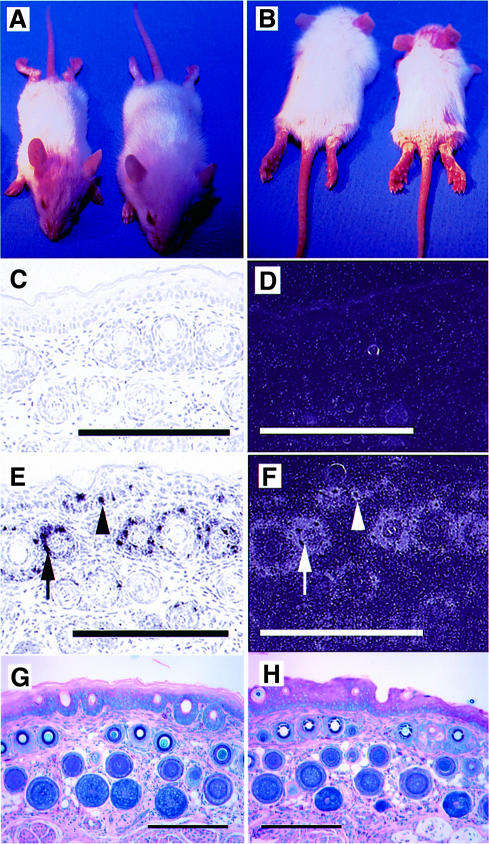
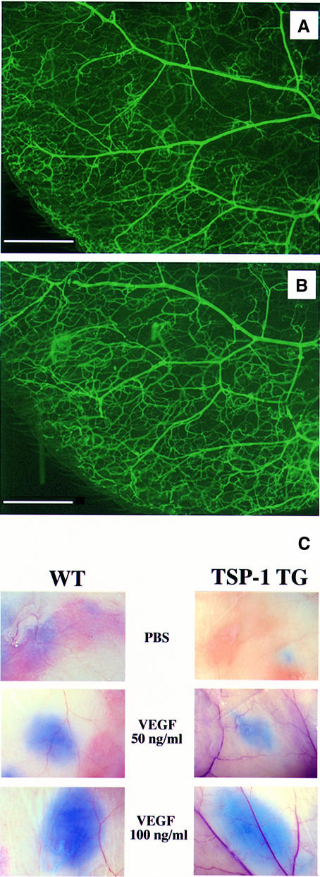
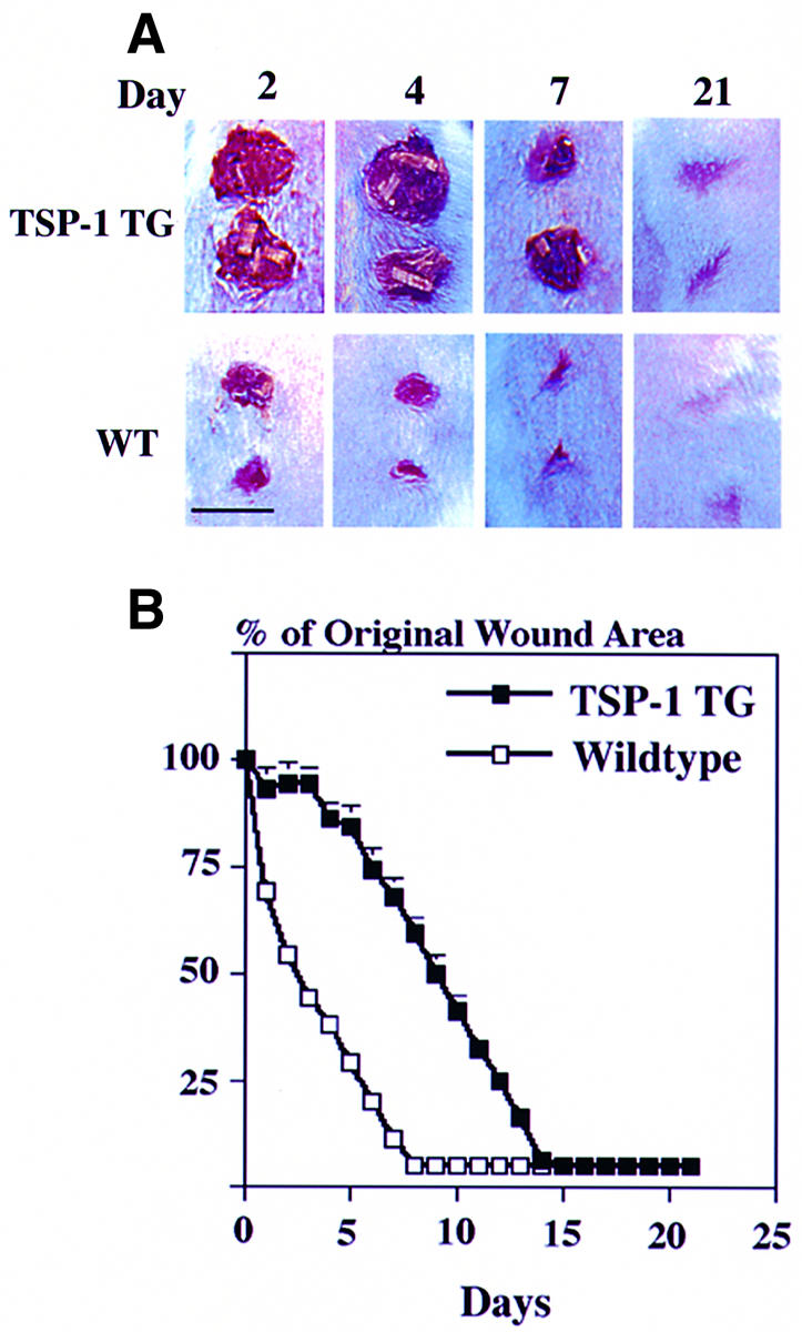
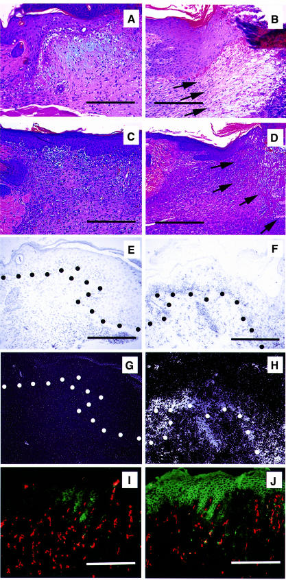
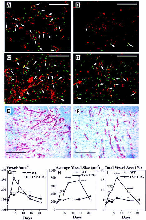
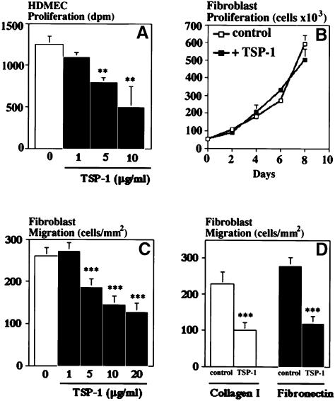
The function of the endogenous angiogenesis inhibitor thrombospondin-1 (TSP-1) in tissue repair has remained controversial. We established transgenic mice with targeted overexpression of TSP-1 in the skin, using a keratin 14 expression cassette. TSP-1 transgenic mice were healthy and fertile, and did not show any major abnormalities of normal skin vascularity, cutaneous vascular architecture, or microvascular permeability. However, healing of full-thickness skin wounds was greatly delayed in TSP-1 transgenic mice and was associated with reduced granulation tissue formation and highly diminished wound angiogenesis. Moreover, TSP-1 potently inhibited fibroblast migration in vivo and in vitro. These findings demonstrate that TSP-1 preferentially interfered with wound healing-associated angiogenesis, rather than with the angiogenesis associated with normal development and skin homeostasis, and suggest that therapeutic application of angiogenesis inhibitors might potentially be associated with impaired wound vascularization and tissue repair.


Figures

None
Fig. 1. (A) Schematic representation of the K14–TSP-1 transgene construct. A 3.6 kb human TSP-1 cDNA fragment was ligated to the BamHI restriction site of the keratin 14 promoter cassette. (B) Overexpression of TSP-1 mRNA in the dorsal and ventral skin of 3-week-old K14–TSP-1 transgenic mice was confirmed by northern blot analysis. TSP-1 mRNA expression was not increased above wild-type (WT) levels in the liver of TSP-1 transgenic mice. Hybridization with a murine β-actin probe served as a control for equal loading. (C) Western blot analysis of skin lysates demonstrates the presence of the intact, 180 kDa TSP-1 protein in TSP-1 transgenic skin. TSP-1, natural human TSP-1. (D) Strong TSP-1 mRNA expression levels in keratinocytes isolated from TSP-1 transgenic mice. TSP-1 expression levels were higher under low Ca2+ conditions than under high Ca2+ conditions that favor keratinocyte differentiation. (E) Western blot analysis confirmed efficient secretion of transgenic TSP-1 protein into culture supernatants and increased protein expression in cell lysates obtained from transgenic keratinocytes, as compared with wild-type (WT) controls.
None
Fig. 2. (A and B) Twelve-day-old TSP-1 transgenic mice (right) were smaller than their wild-type littermates and showed a delayed first hair cycle. TSP-1 transgenic mice were phenotypically indistinguishable from their wild-type littermates after 4 weeks of age. Little TSP-1 mRNA expression was detected by in situ hybridization in wild-type skin (C and D), mainly restricted to follicular keratinocytes of the outer root sheath and to blood vessels. (E and F) Targeted overexpression of the K14–TSP-1 transgene in the basal keratinocyte layer (arrowheads) and in the outer root sheath keratinocytes of hair follicles (arrows) was confirmed by in situ hybridization. Bar: 250 µm. Giemsa staining of the tail skin of adult 7-week-old TSP-1 transgenic mice (H) showed no differences in skin structure as compared with wild-type mice (G). Bar: 250 µm.
None
Fig. 3. Comparable branching pattern and structure of cutaneous blood vessels in adult wild-type mice (A) and TSP-1 transgenic mice (B), visualized in whole-mount preparations of mouse ears after perfusion with fluorescein-labeled L.esculentum lectin. Bar: 1 mm. (C) Microvascular permeability of skin vessels was not altered in TSP-1 transgenic mice (right panels) when compared with wild-type littermates (left panels) in a modified Miles assay. Comparable extravasation of intravenously injected Evans blue was observed after intradermal injection of recombinant VEGF (50 and 100 ng/ml). Injection of PBS served as a control for baseline permeability levels.
None
Fig. 4. Delayed closure of full-thickness wounds in TSP-1 transgenic mice as compared with wild-type (WT) littermates. (A) At 24 h after injury, wounds were covered by a dry scab, which remained adherent until day 10 in TSP-1 transgenic mice and until day 5–7 in control mice. Bar: 6 mm. (B) A >50% reduction in wound area was observed 3 days after wounding in wild-type mice (n = 30) but only after 9 days in TSP-1 transgenic mice (n = 30) (p <0.001). Complete wound closure occurred after 8 days in wild-type mice and after 14 days in TSP-1 transgenic mice.
None
Fig. 5. Impaired granulation tissue formation in cutaneous wounds of TSP-1 transgenic mice. In wild-type mice, the wound bed was completely filled with granulation tissue 3 days after wounding (A), and was completely covered by newly formed epidermis after 7 days (C). In contrast, only minor granulation tissue infiltration from the wound margins was observed in TSP-1 transgenic mice after 3 days (B), and by day 7 the wound bed was still only partially filled with granulation tissue (D). Arrows indicate the front line of granulation tissue invading the wound bed. (A–D) Hematoxylin–eosin stain. Magnification: 96×. Bar: 250 µm. (EHIn situ hybridization confirmed high levels of TSP-1 mRNA expression in transgenic epidermal keratinocytes at day 7 after wounding (F and H), as compared with low TSP-1 mRNA expression in wild-type mice (E and G). Dots indicate the epidermal–dermal junction. Magnification: 192×. Bar: 250 µm. (I and J) Immunofluorescence staining of TSP-1 in 7-day-old wounds reveals strong TSP-1 protein expression in the neoepidermis of TSP-1 transgenic mice (J), as compared with low levels of expression in wild-type wounds (I). Little or no staining of non-resident cells is visible. Vessels are depicted in red (CD31 stain). Magnification: 96×. Bar: 250 µm.
None
Fig. 6. Pronounced inhibition of vascular endothelial cell proliferation and neovascularization in granulation tissue of wounds from TSP-1 transgenic mice at 2 days (B) and 4 days (D) after injury, as compared with numerous proliferating endothelial cells in wild-type mice at day 2 (A) and day 4 (C). Immunofluorescence staining demonstrates CD31-stained blood vessels (red), BrdU-labeled proliferating non-endothelial cells (green) and CD31/BrdU double-stained proliferating vascular endothelial cells (yellow, arrows). Staining for CD31 demonstrated reduced neovascularization of 7-day-old granulation tissue in TSP-1 transgenic mice (F), as compared with wild-type mice (E). Bar: 250 µm. (GI) Computer-assisted morphometric analysis of CD31-stained wound sections revealed a significantly (p <0.01) reduced microvascular density in granulation tissue of TSP-1 transgenic mice (G), as compared with wild-type (WT) controls. (H) Inhibition of vessel size increases through day 14 after injury in granulation tissue of TSP-1 transgenic mice. (I) A strong increase in the total area covered by blood vessels was detected in granulation tissue in wild-type mice, but not in TSP-1 transgenic mice (p <0.001). CD31-stained blood vessels were evaluated in granulation tissue in corresponding 10× fields of five different wounds per time point. Data are expressed as mean ± SEM. **p <0.01; ***p <0.001.
None
Fig. 7. (A) Dose-dependent inhibition of human dermal microvascular endothelial cell (HDMEC) proliferation by TSP-1 in the presence of 20 ng/ml VEGF. (B) Absence of fibroblast growth modulation by added TSP-1 (20 µg/ml), as compared with untreated controls. (C) TSP-1 dose-dependently inhibited in vitro migration of human dermal fibroblasts (p <0.001) towards a collagen type I matrix. (D) Comparable inhibition of fibroblast migration on collagen type I or fibronectin matrices. Transwell migration chamber assay; TSP-1 concentration: 20 µg/ml. Results are the mean ± SD of two independent experiments. **p <0.01; ***p <0.001.

References

    1. Adams J.C. (1997) Thrombospondin-1. Int. J. Biochem. Cell Biol., 29, 861–865. - PubMed
    1. Ando Y. and Jensen,P. (1996) Protein kinase C mediates up-regulation of urokinase and its receptor in the migrating keratinocytes of wounded cultures, but urokinase is not required for movement across a substratum in vitro. J. Cell Physiol., 167, 500–511. - PubMed
    1. Arnold F. and West,D.C. (1991) Angiogenesis in wound healing. Pharmacol. Ther., 52, 407–422. - PubMed
    1. Beer H.-D., Longaker,M.T. and Werner,S. (1997) Reduced expression of PDGF and PDGF receptors during impaired wound healing. J. Invest. Dermatol., 109, 132–138. - PubMed
    1. Border W.A. and Noble,N.A. (1994) Transforming growth factor β in tissue fibrosis. N. Engl. J. Med., 331, 1286–1292. - PubMed

Publication types