. 2000 Jul 3;19(13):3314-24.
doi: 10.1093/emboj/19.13.3314.

BMP-2 antagonists emerge from alterations in the low-affinity binding epitope for receptor BMPR-II

Affiliations

BMP-2 antagonists emerge from alterations in the low-affinity binding epitope for receptor BMPR-II

T Kirsch et al. EMBO J. .

Abstract

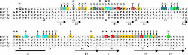
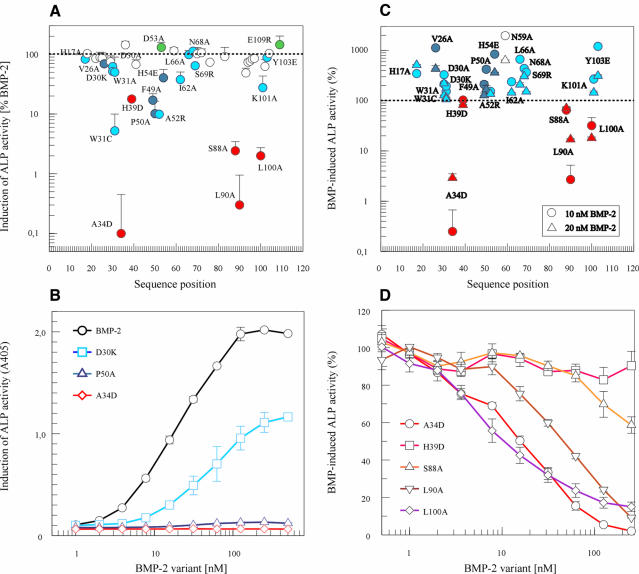
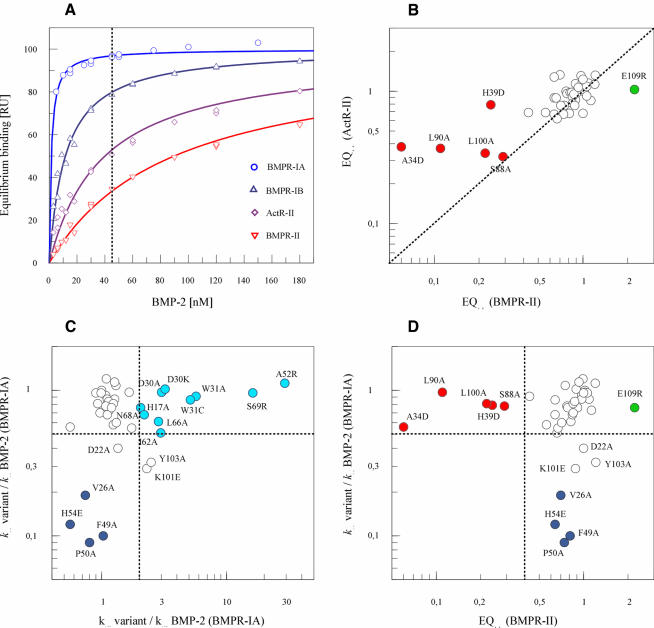
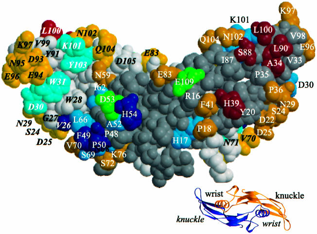
Bone morphogenetic protein-2 (BMP-2) induces bone formation and regeneration in adult vertebrates and regulates important developmental processes in all animals. BMP-2 is a homodimeric cysteine knot protein that, as a member of the transforming growth factor-beta (TGF-beta) superfamily, signals by oligomerizing type I and type II receptor serine-kinases in the cell membrane. The binding epitopes of BMP-2 for BMPR-IA (type I) and BMPR-II or ActR-II (type II) were characterized using BMP-2 mutant proteins for analysis of interactions with receptor ectodomains. A large epitope 1 for high-affinity BMPR-IA binding was detected spanning the interface of the BMP-2 dimer. A smaller epitope 2 for the low-affinity binding of BMPR-II was found to be assembled by determinants of a single monomer. Symmetry-related pairs of the two juxtaposed epitopes occur near the BMP-2 poles. Mutations in both epitopes yielded variants with reduced biological activity in C2C12 cells; however, only epitope 2 variants behaved as antagonists partially or completely inhibiting BMP-2 activity. These findings provide a framework for the molecular description of receptor recognition and activation in the BMP/TGF-beta superfamily.


Figures

None
Fig. 1. BMP-2 residues substituted in this study. Variant BMP-2 proteins with reduced binding affinity for the type II receptor BMPR-II are indicated by the red colour of the substituted position. Altered binding affinities for the type I receptor BMPR-IA due to either a decreased association rate or an increased dissociation rate constant are indicated by dark and light blue of the respective substituted positions. Green indicates positions determining superagonist activity. Yellow positions indicate no measurable alterations in function of the respective variants. Variants substituted at the positions coloured grey could not be isolated in a purity or in amounts sufficient for functional analysis. The structure-based amino acid sequence alignment of BMP-2, BMP-7, TGF-β1 and TGF-β2 as well as the location of secondary structure elements as β-sheets (β1–β9) and α-helix (α3) was adapted from Scheufler et al. (1999). The numbering is according to the BMP-2 sequence.
None
Fig. 2. Biological activity and inhibitory properties of BMP-2 variants. (A) Alkaline phosphatase (ALP) activity was measured after incubation with 250 nM BMP-2 or BMP-2 variant. The response elicited by each variant was calculated as a percentage of the BMP-2 response. Values represent the mean value (±SD) of four measurements. Variants with red symbols are altered in BMPR-II interaction, as shown in Figure 3. Dark or light blue symbols indicate variants with altered association or dissociation rate constants, respectively, for binding to the BMPR-IA receptor chain. (B) Dose-dependent induction of ALP activity in serum-starved C2C12 cells is shown for BMP-2 (circles, black) and for BMP-2 variants D30K (squares, light blue), P50A (triangles, dark blue) and A34D (diamonds, red). (The background absorption at 405 nm of 0.08 ± 0.02 was not subtracted to indicate the signal-to-noise ratio.) (C) The inhibition of induction of ALP activity was determined in serum-starved C2C12 cells after incubation with 250 nM BMP-2 variant in the presence of 10 (circles) or 20 nM (triangles) BMP-2. The response obtained in the presence of BMP-2 alone is indicated by the dotted line and was taken as 100%. Data points represent the mean ± SD of four measurements. Variants with red symbols are altered in BMPR-II interaction. Light or dark blue symbols indicate variants with altered association or dissociation rate constants, respectively for the binding to the BMPR-IA receptor chain. (D) Inhibition of BMP-2 activity (10 nM BMP-2) by increasing doses of putative antagonistic/partial agonistic BMP-2 variants in serum-starved C2C12 cells. Dose–response curves of the variants A34D (circles), H39D (squares), S88A (upright triangles), L90A (inverted triangles) and L100A (diamonds) in the presence of 10 nM BMP-2 were obtained after incubation of the cells for 3 days and analysis of the induced ALP activity.
None
Fig. 3. Interaction of BMP-2 variants with type I (BMPR-IA) or type II (BMPR-II, ActR-II) receptor ectodomains. The association (kon) and dissociation rate constants (koff) for the interaction of a BMP-2 variant at 15, 30 and 45 nM with immobilized BMPR-IA receptor ectodomain were evaluated. Equilibrium binding (EQ45) to the immobilized BMPR-II or ActR-II receptor ectodomain was measured at 45 nM variant. All values were normalized by taking the koff, kon and EQ45 values of BMP-2 as unity. (A) Equilibrium binding at increasing concentrations of BMP-2 to the type I receptors BMPR-IA (circles, light blue) and BMPR-IB (upright triangles, dark blue) as well as to the type II receptors ActR-II (diamonds, magenta) and BMPR-II (inverted triangles, red). (B) Binding affinity of BMP-2 variants for type II receptors BMPR-II or ActR-II. Equilibrium binding during biosensor analysis of 45 nM variant (EQ45) to BMPR-II is plotted versus the binding to ActR-II. (C) Plot of the association rate constants (kon) versus the dissociation rate constants (koff) for the interaction of BMP-2 variants with the BMPR-IA receptor. Variants specifically altered in their kon rates are indicated by dark blue symbols, and those with specifically altered koff by light blue symbols. (D) Plot of the association rate constant (kon) for BMPR-IA binding versus the equilibrium binding (EQ45) to BMPR-II of the same set of variants as in (C). Because both the association rate constant and equilibrium binding are dependent on the concentration of the BMP-2 variant, specific (and concentration-independent) alterations become apparent. (see for example K101E and Y103A for correlation of koff and EQ45). Variants with a specific decrease in equilibrium binding are marked in red.
None
Fig. 4. Residues determining type I (wrist epitope) or type II receptor binding (knuckle epitope) are indicated on the space-filling model of BMP-2. Residues coloured red determine BMPR-II ectodomain binding. Substitutions of residues coloured blue result in altered BMPR-IA receptor binding either by increasing the dissociation rate constant koff (light blue) or by decreasing the association rate constant kon (dark blue). Residues coloured green determine enhanced receptor binding. In the space-filling model, the 2-fold axis of BMP-2 runs in the plane of the figure. The residues of one of the two monomers are indicated by bold italic letters. The residues of the other monomer are coloured with a darker hue. The small ribbon model presents a view of BMP-2 along the 2-fold axis and shows the location of the wrist and knuckle epitopes.

Similar articles

Cited by

References

    1. Black J. (1989) Drugs from emasculated hormones: the principle of syntopic antagonism. Science, 245, 486–493. - PubMed
    1. Blessing M., Schirmacher,P. and Kaiser,S. (1996) Overexpression of bone morphogenetic protein-6 (BMP-6) in the epidermis of transgenic mice: inhibition or stimulation of proliferation depending on the pattern of transgene expression and formation of psoriatic lesions. J. Cell Biol., 135, 227–239. - PMC - PubMed
    1. Chalaux E., Lopez Rovira,T., Rosa,J.L., Bartrons,R. and Ventura,F. (1998) JunB is involved in the inhibition of myogenic differentiation by bone morphogenetic protein-2. J. Biol. Chem., 273, 537–543. - PubMed
    1. Chen Y., Riese,M.J., Killinger,M.A. and Hoffmann,F.M. (1998) A genetic screen for modifiers of Drosophila decapentaplegic signaling identifies mutations in punt, Mothers against dpp and the BMP-7 homologue, 60A. Development, 125, 1759–1768. - PubMed
    1. Cunningham B.C. and Wells,J.A. (1993) Comparison of a structural and a functional epitope [published erratum appears in J. Mol. Biol.,237, 513]. J. Mol. Biol., 234, 554–563. - PubMed

Publication types

MeSH terms

Substances