. 2000 Jul 3;19(13):3325-36.
doi: 10.1093/emboj/19.13.3325.

The apoptotic signaling pathway activated by Toll-like receptor-2

Affiliations

The apoptotic signaling pathway activated by Toll-like receptor-2

A O Aliprantis et al. EMBO J. .

Abstract

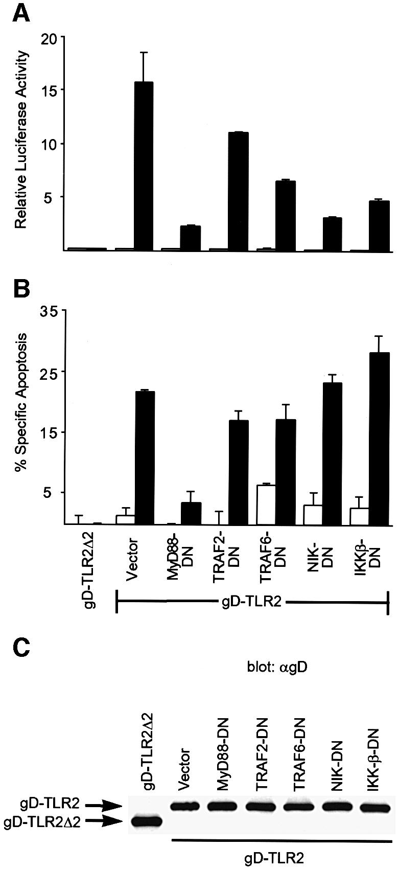
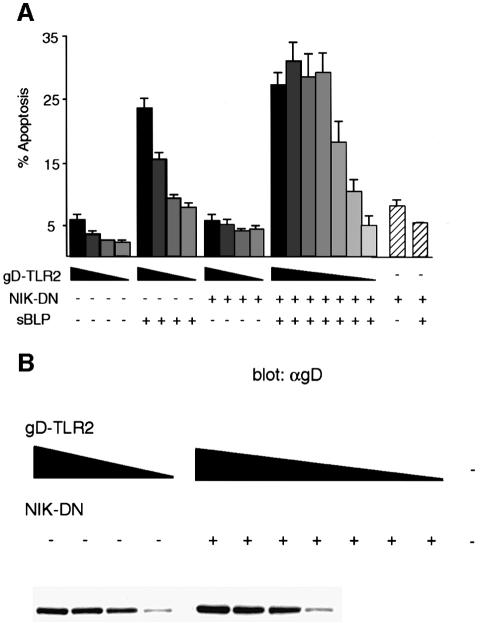
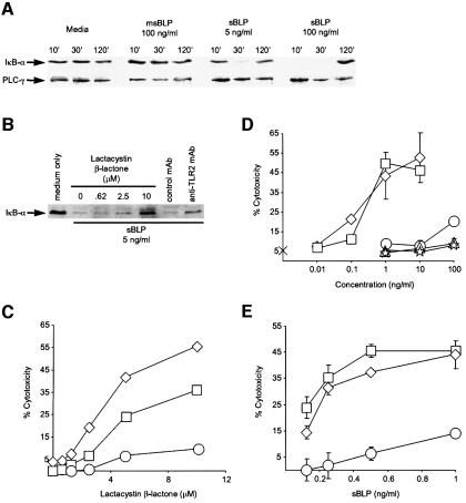
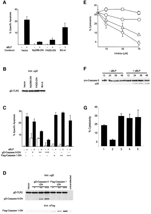
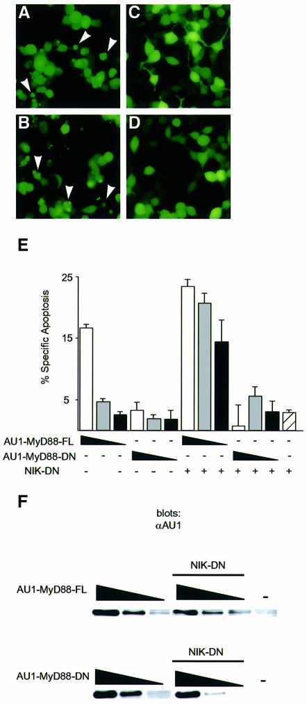
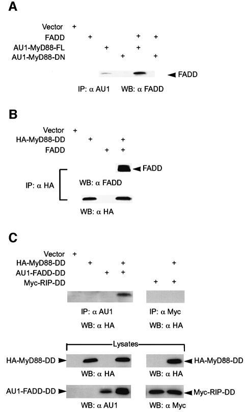
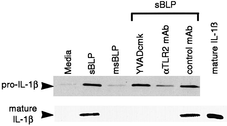
The innate immune system uses Toll family receptors to signal for the presence of microbes and initiate host defense. Bacterial lipoproteins (BLPs), which are expressed by all bacteria, are potent activators of Toll-like receptor-2 (TLR2). Here we show that the adaptor molecule, myeloid differentiation factor 88 (MyD88), mediates both apoptosis and nuclear factor-kappaB (NF-kappaB) activation by BLP-stimulated TLR2. Inhibition of the NF-kappaB pathway downstream of MyD88 potentiates apoptosis, indicating that these two pathways bifurcate at the level of MyD88. TLR2 signals for apoptosis through MyD88 via a pathway involving Fas-associated death domain protein (FADD) and caspase 8. Moreover, MyD88 binds FADD and is sufficient to induce apoptosis. These data indicate that TLR2 is a novel 'death receptor' that engages the apoptotic machinery without a conventional cytoplasmic death domain. Through TLR2, BLP induces the synthesis of the precursor of the pro-inflammatory cytokine interleukin-1beta (IL-1beta). Interestingly, BLP also activates caspase 1 through TLR2, resulting in proteolysis and secretion of mature IL-1beta. These results indicate that caspase activation is an innate immune response to microbial pathogens, culminating in apoptosis and cytokine production.


Figures

None
Fig. 1. MyD88 is a component of the NF-κB and apoptotic pathways initiated by BLP through hTLR2. (A and B) 293 cells were transiently co-transfected with equivalent amounts (0.25 µg) of expression plasmids encoding gD-TLR2 or gD-TLR2Δ2 and the indicated dominant negatives. An NF-κB-regulated luciferase reporter plasmid was also transfected. At 24 h post-transfection, the cells were incubated in medium alone (white bars) or in medium with 1 µg/ml sBLP (black bars). The induction of NF-κB-dependent transcription (A) was determined. Apoptosis (B) was measured by TUNEL analysis. Data in (B) are reported as the percentage specific apoptosis relative to cells transfected with gD-TLR2Δ2 without sBLP. Results are the average ± SD of two independent samples. (C) Anti-gD western blot of lysates prepared from cells transfected as in (A) and (B).
None
Fig. 2. Inhibition of the NF-κB pathway facilitates TLR2-mediated apoptosis. (A) 293 cells were transiently transfected with 0.25, 0.1, 0.04, 0.01 and, in addition for co-transfections with NIK-DN, 0.002, 0.0004 and 0.00008 µg of an expression plasmid encoding gD-TLR2 indicated in a gray gradient from black (0.25 µg) to white (0.00008 µg). Where indicated, the cells were also transfected with 0.25 µg of an expression plasmid encoding NIK-DN. Control cells transfected with vector DNA only are indicated with hatched bars. At 24 h post-transfection, the cells were incubated with or without 1 µg/ml sBLP as indicated and apoptosis was determined by TUNEL staining. (B) Anti-gD western blot of lysates prepared from cells transfected as in (A). Cells transfected with 0.002 µg or less had undetectable levels of gD-TLR2.
None
Fig. 3. Inhibition of IκB-α degradation augments TLR2-mediated cell death in THP-1 cells. (A) sBLP induces the degradation of IκB-α in THP-1 cells. THP-1 cells were incubated with medium, msBLP (100 ng/ml) or sBLP (5 or 100 ng/ml) for the indicated times and analyzed by western blotting for IκB-α and for PLCγ, as a loading control. (B) A proteasome inhibitor and an anti-TLR2 blocking mAb inhibit IκB-α degradation in sBLP-treated THP-1 cells. THP-1 cells were pre-incubated with either medium alone, the indicated concentrations of lactacystin β-lactone, 25 µg/ml anti-TLR2 mAb 2392 or an isotype control mAb. sBLP was added to a final concentration of 5 ng/ml. After 30 min, the cells were analyzed as in (A). (C) Lactacystin β-lactone facilitates sBLP-mediated apoptosis in THP-1 cells. THP-1 cells were pre-incubated with the indicated concentrations of lactacystin β-lactone. Either medium alone (circles) or medium with sBLP was added to yield final sBLP concentrations of 2.5 ng/ml (diamonds) or 0.25 ng/ml (squares). (D) sBLP and MLP, but not Pam3Cys, msBLP or LPS, kill lactacystin β-lactone-treated THP-1 cells. THP-1 cells were pre-incubated for 1 h with 5 µM lactacystin β-lactone. Medium only (cross) or medium with either sBLP (diamonds), MLP (squares), Pam3Cys (circles), msBLP (triangles) or LPS (stars) was added to yield the indicated final concentrations. (E) sBLP-induced death of THP-1 cells pre-treated with lactacystin β-lactone is dependent on TLR2. THP-1 cells were pre-incubated for 1 h with 10 µM lactacystin β-lactone in either medium alone (diamonds) or medium with 25 µg/ml anti-TLR2 mAb 2392 (circles) or an isotype control mAb (squares). sBLP was added to yield the indicated final concentrations. Some standard deviations in (C), (D) and (E) are within the limits of the data points.
None
Fig. 4. TLR2-induced apoptosis proceeds through a FADD/caspase 8 pathway. (A) TLR2-mediated apoptosis is inhibited by FADD-DN but not by Bcl-xl. 293 cells were transiently co-transfected with 0.25 µg of an expression plasmid encoding gD-TLR2 and 0.3 µg of expression plasmids encoding the indicated dominant negatives or Bcl-xl. At 24 h post-transfection, the cells were incubated with or without 1 µg/ml sBLP as indicated and apoptosis was determined by TUNEL staining. (B) Anti-gD western blot of lysates prepared from cells transfected as in (A) indicating equivalent expression of gD-TLR2 among transfections. (C) TLR2-mediated apoptosis is inhibited by a catalytically inactive mutant of caspase 8. 293 cells were transiently co-transfected with 0.15 µg of an expression plasmid encoding gD-TLR2 and either 0.05 (+), 0.15 (++) or 0.45 µg (+++) of expression plasmids encoding gD-caspase 8-DN or Flag-caspase 1-DN. Apoptosis was induced and quantified as in (A). Data in (A) and (C) are reported as the percentage specific apoptosis relative to cells transfected with gD-TLR2 without sBLP. (D) Anti-gD (upper panel) and anti-Flag (lower panel) western blots of lysates prepared from cells transfected as in (C). (E) sBLP-mediated cell death of THP-1 cells is attenuated by a peptide inhibitor of caspase 8. THP-1 cells were pre-incubated with medium containing 75 µg/ml cycloheximide without (star) or with the indicated concentrations of z-FA-fmk (squares), Ac-YVAD-cmk (circles), z-IETD-fmk (triangles) or z-VAD-fmk (diamonds). sBLP was added to a final concentration of 2 ng/ml. Some standard deviations are within the limits of the data points. (F) Anti-casapse-8 western blots of lysates prepared from 293 cells co-transfected with gD-TLR2 and NIK-DN and incubated for the indicated times (h) with sBLP (1 µg/ml). Pro-caspase 8 (p55) and the p28 proteolytic intermediate are shown. (G) sBLP-mediated cell death is not mediated through TNF or Fas. THP-1 cells were pre-incubated for 4.5 h with medium only (1) or with 5 µg/ml anti-TLR2 mAb 2392 (2), an isotype-matched control mAb (3), anti-Fas (4) or anti-TNF mAb (5) and then with 100 pg/ml sBLP. Cytotoxicity was assayed by LDH release.
None
Fig. 5. MyD88-FL, but not MyD88-DN, induces apoptosis in 293 cells. (A–D) 293 cells were transiently transfected with 0.25 µg of expression plasmids encoding AU1-MyD88-FL (A and B) or AU1-MyD88-DN (C and D). In (B) and (D), 0.25 µg of an expression plasmid encoding NIK-DN was co-transfected. A plasmid encoding GFP (0.025 µg) was included in the transfection to label transfected cells. GFP-positive cells were photographed 48 h post-transfection. Arrowheads indicate cells with apoptotic morphology. (E) Inhibition of the NF-κB pathway facilitates MyD88-mediated apoptosis. 293 cells were transiently transfected with 0.25 (white bars), 0.1 (gray), 0.04 (black) or 0 µg (diagonal lines) of expression plasmids encoding AU1-MyD88-FL or AU1-MyD88-DN. Where indicated, 0.25 µg of an expression plasmid encoding NIK-DN was co-transfected. A plasmid encoding GFP (0.025 µg) was included to label transfected cells. At 60 h post-transfection, apoptosis was determined by annexin V staining. The data are reported as the percentage specific apoptosis in the GFP-positive (transfected) population relative to cells transfected with vector DNA. A specific increase in apoptosis was only detected in the GFP-positive population. (F) Anti-AU1 western blot of lysates prepared from cells transfected as in (E).
None
Fig. 6. MyD88 associates with FADD through its DD–DD interaction. (A) 293 cells were transiently transfected with expression plasmids (1 µg each) encoding FADD, AU1-MyD88-FL or AU1-MyD88-DN alone, or in combination as indicated. At 24 h post-transfection, cell lysates were prepared, and co-immunoprecipitation (IP) and western blotting were performed using antisera as indicated. (B) 293 cells were transiently transfected with expression plasmids (1 µg each) encoding FADD and/or HA epitope-tagged MyD88-DD. The experiment was performed as in (A) except that an anti-HA mAb was used for the IP. (C) 293 cells were transiently transfected with expression plasmids (1 µg each) encoding the death domains (DDs) of MyD88, FADD or RIP alone or in combination. At 24 h post-transfection, cell lysates were prepared, and immunoprecipitation (IP) and western blotting (WB) were performed using the indicated antibodies.
None
Fig. 7. TLR2 mediates pro-IL-1β synthesis and caspase 1 activation in THP-1 cells treated with sBLP. THP-1 cells were pre-treated for 6 h with 32 nM PMA in the indicated conditions. Where shown, cells were pre-incubated with 10 µM Ac-YVAD-cmk, 25 µg/ml anti-TLR2 mAb 2392 or isotype control mAb and then treated with sBLP at 4 ng/ml. After 3 h, cell lysates were resolved by SDS–PAGE and blotted for pro-IL-1β. Culture supernatants were collected, resolved by SDS–PAGE and blotted for mature IL-1β. As a control for the molecular weight of mature IL-1β, 1 ng of recombinant mature IL-1β was resolved and blotted in parallel.

References

    1. Adachi O., Kawai,T., Takeda,K., Matsumoto,M., Tsutsui,H., Sakagami,M., Nakanishi,K. and Akira,S. (1998) Targeted disruption of the MyD88 gene results in loss of IL-1- and IL-18-mediated function. Immunity, 9, 143–150. - PubMed
    1. Aliprantis A.O., Yang,R.B., Mark,M.R., Suggett,S., Devaux,B., Radolf,J.D., Klimpel,G.R., Godowski,P. and Zychlinsky,A. (1999) Cell activation and apoptosis by bacterial lipoproteins through Toll-like receptor-2. Science, 285, 736–739. - PubMed
    1. Ashkenazi A. and Dixit,V.M. (1998) Death receptors: signaling and modulation. Science, 281, 1305–1308. - PubMed
    1. Baeuerle P.A. (1998) IκB–NF-κB structures: at the interface of inflammation control. Cell, 95, 729–731. - PubMed
    1. Baeuerle P.A. and Baichwal,V.R. (1997) NF-κ B as a frequent target for immunosuppressive and anti-inflammatory molecules. Adv. Immunol., 65, 111–137. - PubMed

Publication types

MeSH terms

Substances