. 2000 Jul 3;19(13):3349-57.
doi: 10.1093/emboj/19.13.3349.

Circadian clock-protein expression in cyanobacteria: rhythms and phase setting

Affiliations

Circadian clock-protein expression in cyanobacteria: rhythms and phase setting

Y Xu et al. EMBO J. .

Abstract

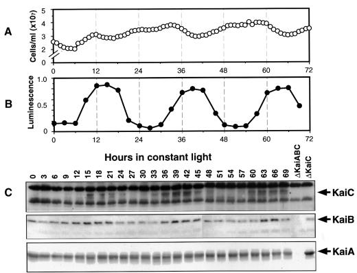
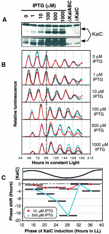
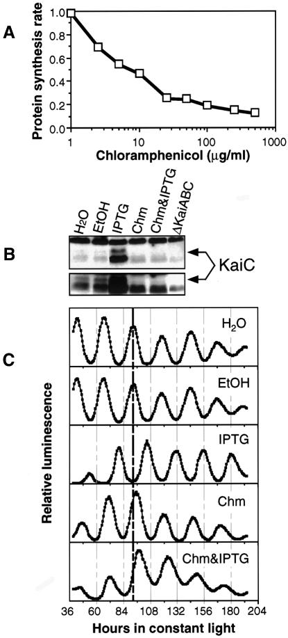
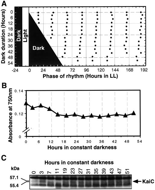
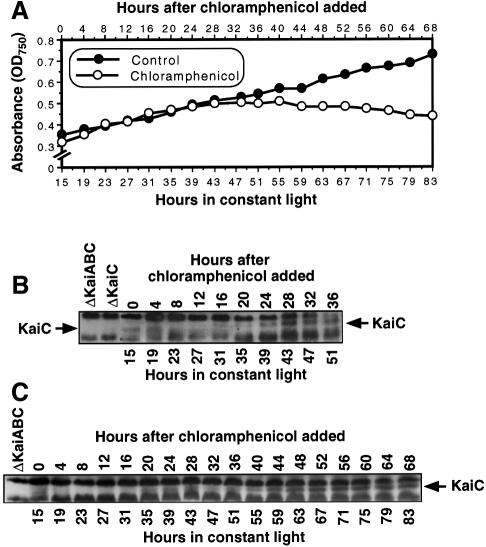
The cyanobacterial gene cluster kaiABC encodes three essential circadian clock proteins: KaiA, KaiB and KaiC. The KaiB and KaiC protein levels are robustly rhythmical, whereas the KaiA protein abundance undergoes little if any circadian oscillation in constant light. The level of the KaiC protein is crucial for correct functioning of the clock because induction of the protein at phases when the protein level is normally low elicits phase resetting. Titration of the effects of the inducer upon phase resetting versus KaiC level shows a direct correlation between induction of the KaiC protein within the physiological range and significant phase shifting. The protein synthesis inhibitor chloramphenicol prevents the induction of KaiC and blocks phase shifting. When the metabolism is repressed by either translational inhibition or constant darkness, the rhythm of KaiC abundance persists; therefore, clock protein expression has a preferred status under a variety of conditions. These data indicate that rhythmic expression of KaiC appears to be a crucial component of clock precession in cyanobacteria.


Figures

None
Fig. 1. Rhythms of Kai proteins for 3 days in LL from log-phase cultures that are continuously diluted to maintain an approximately equal cell concentration (30°C; light intensity = 125 µE/m2/s; strain = AMC149). In this experiment, the average generation time was 9.45 h. (A) Cell counts of culture showing the approximately equal cell numbers at all phases as in Mori et al. (1996). (B) Luminescence of the culture that reports the circadian rhythm of the psbAI promoter activity. (C) Immunoblots of protein extracts collected at the various phases. The blots were treated with antibodies to KaiC (top strip), KaiB (middle strip) and KaiA (bottom strip). The Kai-specific bands are indicated by the arrows to the right of the panels; note the ΔKaiABC and ΔKaiC lanes that are the controls to indicate non-specific bands.
None
Fig. 2. Dose response of KaiC induction and clock resetting (strain = trcp::kaiC). Cells came from a continuously diluted culture grown as in Figure 1. (A) Induction of KaiC by 6 h treatments of IPTG given at the same CT as in the rhythm experiment shown in (B). The KaiC-specific band is indicated by the arrow to the right of the panel. The exposure time of the upper part is comparable to the exposure of the KaiC blot in Figure 1C, whereas the lower part is the relevant area of the same blot that was exposed longer to visualize the weaker bands. (B) Resetting of the luminscence rhythm by 6 h pulses of various concentrations of IPTG. After a 12 h synchronizing dark pulse, cyanobacterial cultures harboring the trcp::kaiC construct were placed in LL. The 6 h IPTG pulse was begun 21 h after the beginning of LL. Triplicate samples for each concentration are shown in different colors. The heavy vertical dashed line at hour 92 shows the control phase for comparison. (C) Phase-dependent resetting by KaiC induction in the trcp::kaiC strain. Upper part, the rhythm of KaiC abundance abstracted from the data depicted in Figure 1C. Lower part, phase resetting of the luminescence rhythm by IPTG induction of KaiC. Six hour pulses of IPTG at either 10 or 500 µM were administered to liquid cultures of cyanobacteria at different phases in LL. The midpoints of the 6 h pulses are connected by the lines. The phase shifts shown are the averages of duplicates for each point. The phase response curves are plotted monotonically as all-delay phase shifts (Johnson, 1999).
None
Fig. 3. Inhibition of KaiC induction and clock resetting by chloramphenicol in the trcp::kaiC strain. Cells were grown as in Figure 1 until the time of IPTG and/or chloramphenicol addition. (A) Inhibition of total protein synthesis by chloramphenicol. Protein synthetic rate is expressed as percent incorporation (incorporation/uptake) of [35S]l-methionine and [35S]l-cysteine with the control normalized to 1.0 (chloramphenicol concentration expressed as log concentration). Chloramphenicol was dissolved in ethanol; the final concentration of ethanol in all samples was 0.2%. (B) Inhibition of KaiC induction by chloramphenicol assayed by immunoblotting. Concentrations: 50 µg/ml chloramphenicol (Chm) in 0.2% ethanol; 1 mM IPTG (dissolved in dH2O); ethanol, 0.2%. The lower panel is the relevant region of the blot overexposed to visualize the expression level in the control sample. (C) Inhibition of IPTG-induced phase resetting by 50 µg/ml chloramphenicol. Six hour pulses of water, 0.2% ethanol, 1 mM IPTG, chloramphenicol (50 µg/ml in 0.2% ethanol) or IPTG plus chloramphenicol were initiated at hour 21 of LL as in Figure 2B.
None
Fig. 4. The cyanobacterial clock and the KaiC oscillation continues to run in constant darkness in the AMC149 strain. Cells were grown for 4 days in an LD 12:12 cycle until the OD750 was 0.13. At that time, cultures were transferred to DD. (A) The clock continues to run in DD, as assessed by returning samples to LL after various durations of exposure to DD. Shaded areas indicate time of darkness; filled circles indicate the phases of the troughs of the luminescence rhythm during the subsequent free run in LL. (B) Cultures stop growing immediately in DD (measured by OD750). (C) KaiC immunoblot of cultures in DD (note that there is no sample for hour 17). To compare the phasing of the KaiC abundance rhythm in DD with that in LL, note that hour 0 in Figure 4 corresponds to hour 12 in Figure 1 (both are CT12). Molecular weights of the KaiC bands were calculated on the basis of migration.
None
Fig. 5. Chloramphenicol treatment does not set the phase of the circadian clock (strain = AMC149). Cells were grown in batch culture prior to addition of chloramphenicol. Chloramphenicol (50 µg/ml) was applied to cyanobacterial cultures for various durations, then removed. Filled circles indicate the phases of the troughs of the luminescence rhythm after wash-out of chloramphenicol. (A) Chloramphenicol treatment beginning immediately after the synchronizing dark treatment. (B) Chloramphenicol treatment beginning 12 h after the synchronizing dark treatment.
None
Fig. 6. KaiC abundance is still rhythmic in the presence of chloramphenicol. The cell culture for this experiment was a batch culture grown to an OD750 of 0.3–0.4, then 50 µg/ml chloramphenicol was added to cultures 15 h after the onset of LL (strain = AMC149). (A) In the presence of 50 µg/ml chloramphenicol, cell growth and division continue for ∼24 h in chloramphenicol, then stop, whereas the control culture continues to grow (growth measured by OD750). (B) KaiC immunoblot for a culture in chloramphenicol for 36 h (experiment 1). (C) KaiC immunoblot for a culture in chloramphenicol for 68 h (experiment 2).

References

    1. Aoki S., Kondo,T., Wada,H. and Ishiura,M. (1997) Circadian rhythm of the cyanobacterium Synechocystis sp. strain PCC 6803 in the dark. J. Bacteriol., 179, 5751–5755. - PMC - PubMed
    1. Aronson B., Johnson,K., Loros,J.J. and Dunlap,J.C. (1994) Negative feedback defining a circadian clock: autoregulation in the clock gene frequency. Science, 263, 1578–1584. - PubMed
    1. Bustos S.A. and Golden,S.S. (1991) Expression of the psbDII gene in Synechococcus sp. strain PCC 7942 requires sequences downstream of the transcription start site. J. Bacteriol., 173, 7525–7533. - PMC - PubMed
    1. Darlington T.K., Wager-Smith,K., Ceriani,M.F., Staknis,D., Gekakis,N., Steeves,T.D.L., Weitz,C.J., Takahashi,J.S. and Kay,S.A. (1998) Closing the circadian loop: CLOCK-induced transcription of its own inhibitors per and tim. Science, 280, 1599–1603. - PubMed
    1. Doolittle W.F. (1979) The cyanobacterial genome, its expression and the control of that expression. Adv. Microb. Physiol., 20, 1–102. - PubMed

Publication types