. 2000 Jul 3;19(13):3446-57.
doi: 10.1093/emboj/19.13.3446.

Isolation and characterization of Tn7 transposase gain-of-function mutants: a model for transposase activation

Affiliations

Isolation and characterization of Tn7 transposase gain-of-function mutants: a model for transposase activation

F Lu et al. EMBO J. .

Abstract

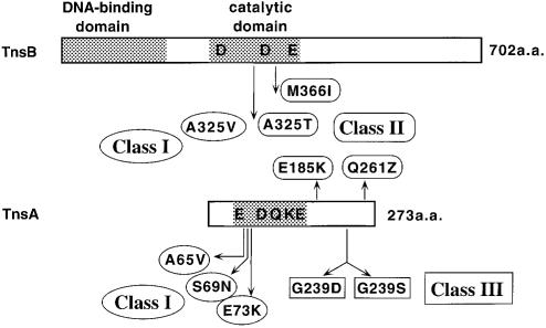
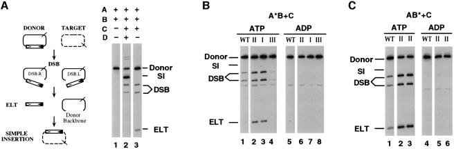
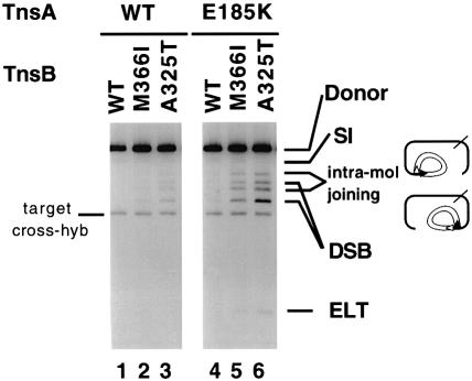
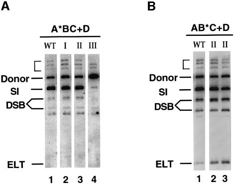
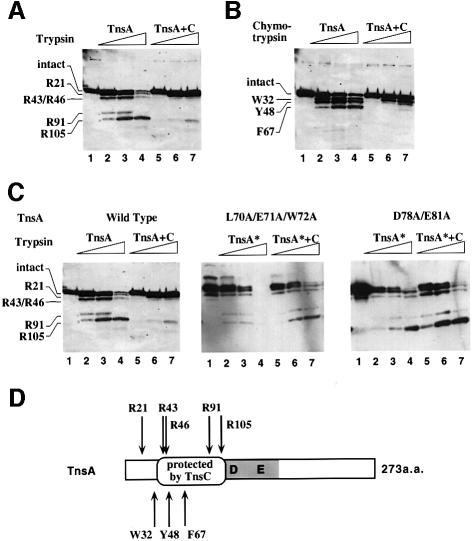
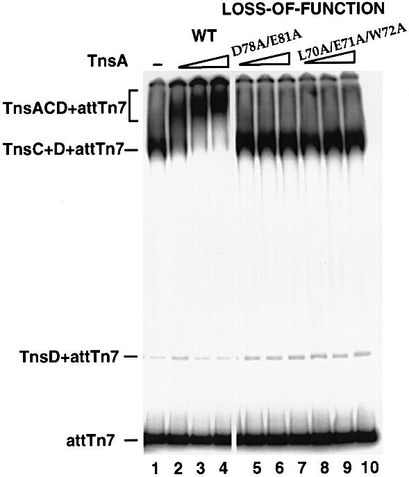
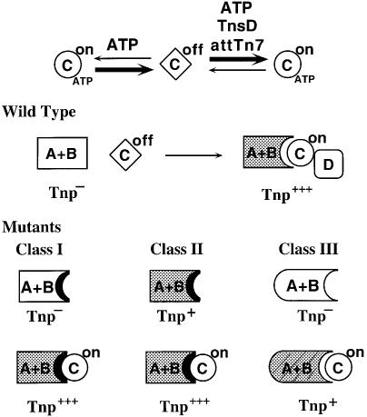
Tn7 transposition has been hypothesized to require a heteromeric transposase formed by two Tn7-encoded proteins, TnsA and TnsB, and accessory proteins that activate the transposase when they are associated with an appropriate target DNA. This study investigates the mechanism of Tn7 transposase activation by isolation and analysis of transposase gain-of-function mutants that are active in the absence of these accessory proteins. This work shows directly that TnsA and TnsB are essential and sufficient components of the Tn7 transposase and also provides insight into the signals that activate the transposase. We also describe a protein-protein interaction between TnsA and TnsC, a regulatory accessory protein, that is likely to be critical for transposase activation.


Figures

None
Fig. 1. Location of the transposase gain-of-function mutations. Schematic illustration of the functional domains of TnsA and TnsB and various point mutations collected in this study. The shaded boxes represent the DNA binding domain of TnsB (R.Sarnovsky and N.L.Craig, unpublished data), the catalytic domain of TnsB (Sarnovsky et al., 1996), and the catalytic domain of TnsA (Sarnovsky et al., 1996; Hickman et al., 2000). The characters ‘D’, ‘E’, ‘K’ and ‘Q’ indicate the relative positions of amino acids implicated in catalysis: D273, D361 and E396 in TnsB; E63, D114, Q130, K132 and E149 in TnsA. The arrows indicate the relative positions of amino acid changes in the transposase gain-of-function mutants. The mutant classes are explained in the text.
None
Fig. 2. Recombination activity of the mutants in TnsABC reaction and the effect of ATP cofactor. (A) The Tn7 transposition pathway is illustrated on the left. Donor DNA (solid line) containing the miniTn7 transposon (rectangle); target DNA (dashed line). The cis-acting recombination sequences at the left and right ends of the transposon are indicated by open and filled triangles, respectively. miniTn7 is excised from the donor DNA by double-strand breaks, through the intermediates DSB.L (double-strand break at the left end), DSB.R (double-strand break at the right end) and ELT (excised linear transposon). ELT then can be inserted into the target DNA to give a simple insertion product (SI). The positions of the NdeI sites used to linearize the plasmid substrates are indicated by slashed lines. A Southern blot of the in vitro transposition reactions is shown on the right. The proteins included in each reaction are indicated above each lane. (B) TnsA*BC reactions: TnsAE185K from class II (lanes 2 and 6), TnsAS69N from class I (lanes 3 and 7) and TnsAG239D from class III (lanes 4 and 8). As indicated, either ADP or ATP was used in the assembly reaction mixture. (C) TnsAB*C reactions: TnsBM366I (lanes 2 and 5) and TnsBA325T (lanes 3 and 6) from class II.
None
Fig. 3. Transposase gain-of-function mutants can support breakage and joining reaction with only TnsA and TnsB. TnsAB reactions with mutant proteins. The assembly step was carried out without either TnsC or TnsD under the conditions specified in Materials and methods except for the additional 20% glycerol. ‘intra-mol joining’ indicates intramolecular joining products where one end of the transposon has been cleaved and subsequently joined within the same donor in the region near to the other end of the transposon (Biery et al., 2000b). The low-level cross-hybridization to the target DNA occasionally observed with this probe is indicated.
None
Fig. 4. Effect of the target selection protein TnsD in vitro. (A) TnsA*BC+D reactions: TnsAS69N from class I (lane 2), TnsAE185K from class II (lane 3) and TnsAG239D from class III (lane 4). The three bands above the donor substrate, indicated with a bracket, are intermediate products resulting from the joining between a partially cleaved donor substrate and the target DNA. (B) TnsAB*C+D reactions: TnsBM366I (lane 2) and TnsBA325T (lane 3) from class I.
None
Fig. 5. Localization of a specific region in TnsA that is involved in direct interaction with TnsC. (A) Protease footprinting of TnsA using trypsin. Lane 1, untreated TnsA; lanes 2–4, TnsA treated with increasing amounts of trypsin (10, 33 and 100 ng); lanes 5–7, TnsA was preincubated with TnsC and then treated with increasing amounts of trypsin (33, 100 and 333 ng). The first N-terminal amino acid of the proteolytic products is labeled. (B) Protease footprinting of TnsA using chymotrypsin. Lane 1, untreated TnsA; lanes 2–4, TnsA treated with chymotrypsin (3.3, 10 and 33 ng); lanes 5–7, TnsA was preincubated with TnsC and then treated with chymotrypsin (10, 33 and 100 ng). (C) Protease footprinting of TnsA mutants. Lane 1, untreated TnsA; lanes 2–4, TnsA treated with increasing amounts of trypsin (10, 33 and 100 ng); lanes 5–7, TnsA preincubated with TnsC and then treated with increasing amounts of trypsin (10, 33 and 100 ng). (D) The protease cleavage sites on TnsA are indicated by arrows (top: trypsin; bottom: chymotrypsin). The region protected by TnsC is marked between amino acids 43 and 105. The shaded box represents the catalytic domain of TnsA (Sarnovsky et al., 1996; Hickman et al., 2000). The characters ‘D’ and ‘E’ indicate the D114 and E149 amino acids implicated in catalysis; other positions—E63, Q130 and K132—are not indicated in this view.
None
Fig. 6. Effect of TnsA loss-of-function mutations on the TnsA–TnsC interaction analyzed by gel mobility shift assay. The DNA substrate contains attTn7 (–52 to +64). TnsC and TnsD were present in all the reactions. Lane 1, no TnsA was added. Lanes 2–4, wild-type TnsA (13, 26 and 40 ng). Lanes 5–10, loss-of-function mutants were added: lanes 5–7, mutant D78A/E81A (13, 26 and 40 ng); lanes 8–10, mutant L70A/E71A/W72A (13, 26 and 40 ng).
None
Fig. 7. Location of TnsA mutations on the structure of TnsA. (A) Ribbons (Carson, 1997) representation of the three-dimensional structure of TnsA. The N-terminal catalytic domain is in blue, the C-terminal helix–turn–helix containing domain is in orange. Class I residues on helix α2 are marked in red, class II residues are in green. The location of the class III mutations is marked on the main chain just upstream of α10. Residues that are essential for catalytic activity (E63, D114 and K132) and the two bound catalytic Mg2+ residues are also shown. (B) The molecular surface of TnsA overlaid to its ribbons trace. Exposed hydrophobic areas are painted green on the surface. Class I residues are marked in red; they are located on helix α2, which forms one of the boundaries that is part of a large hydrophobic patch on the surface. The surface calculations were performed with SPOCK (Christopher, 1998), the ribbon was drawn with Molscript (Kraulis, 1991) and the final figure was rendered with Povray 3.1 (persistence of vision at http://www.povray.org).
None
Fig. 8. Model for Tn7 transposase activation. TnsC is present as two forms: TnsCon (circle) and TnsCoff (rhombus). The equilibrium between the two forms is determined by ATP and targeting cofactors TnsD and attTn7. The wild-type TnsAB transposase by itself is inactive (clear) and is unable to promote transposition (Tnp). TnsAB can be activated (gray) by active TnsC in TnsCATP+TnsD+attTn7 complex and mediate transposition (Tnp+). The impact of mutational changes on the transposase is shown at the bottom and elucidated in the text.

Similar articles

Cited by

References

    1. Arciszewska L.K. and Craig,N.L. (1991) Interaction of the Tn7-encoded transposition protein TnsB with the ends of the transposon. Nucleic Acids Res., 19, 5021–5029. - PMC - PubMed
    1. Arciszewska L.K., McKown,R.L. and Craig,N.L. (1991) Purification of TnsB, a transposition protein that binds to the ends of Tn7. J. Biol. Chem., 266, 21736–21744. - PubMed
    1. Bainton R., Gamas,P. and Craig,N.L. (1991) Tn7 transposition in vitro proceeds through an excised transposon intermediate generated by staggered breaks in DNA. Cell, 65, 805–816. - PubMed
    1. Bainton R.J., Kubo,K.M., Feng,J.-N. and Craig,N.L. (1993) Tn7 transposition: target DNA recognition is mediated by multiple Tn7-encoded proteins in a purified in vitro system. Cell, 72, 931–943. - PubMed
    1. Baker T.A. and Luo,L. (1994) Identification of residues in the Mu transposase essential for catalysis. Proc. Natl Acad. Sci. USA, 91, 6654–6658. - PMC - PubMed

Publication types