. 2000 Jul 3;19(13):3465-74.
doi: 10.1093/emboj/19.13.3465.

Adenovirus-specific translation by displacement of kinase Mnk1 from cap-initiation complex eIF4F

Affiliations

Adenovirus-specific translation by displacement of kinase Mnk1 from cap-initiation complex eIF4F

R Cuesta et al. EMBO J. .

Abstract

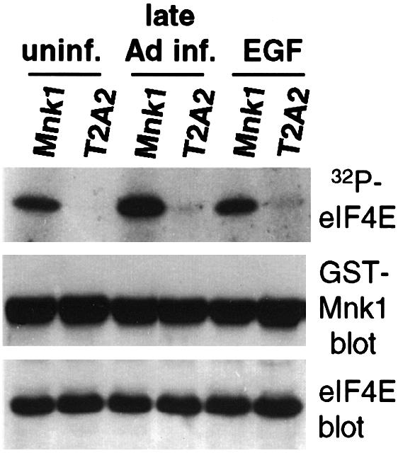
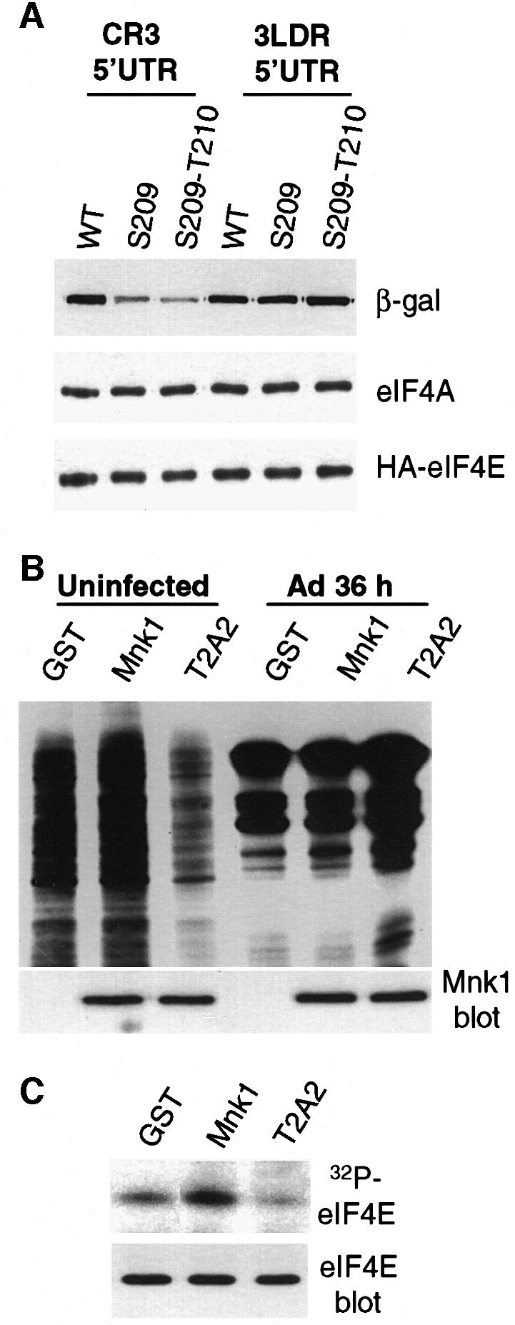
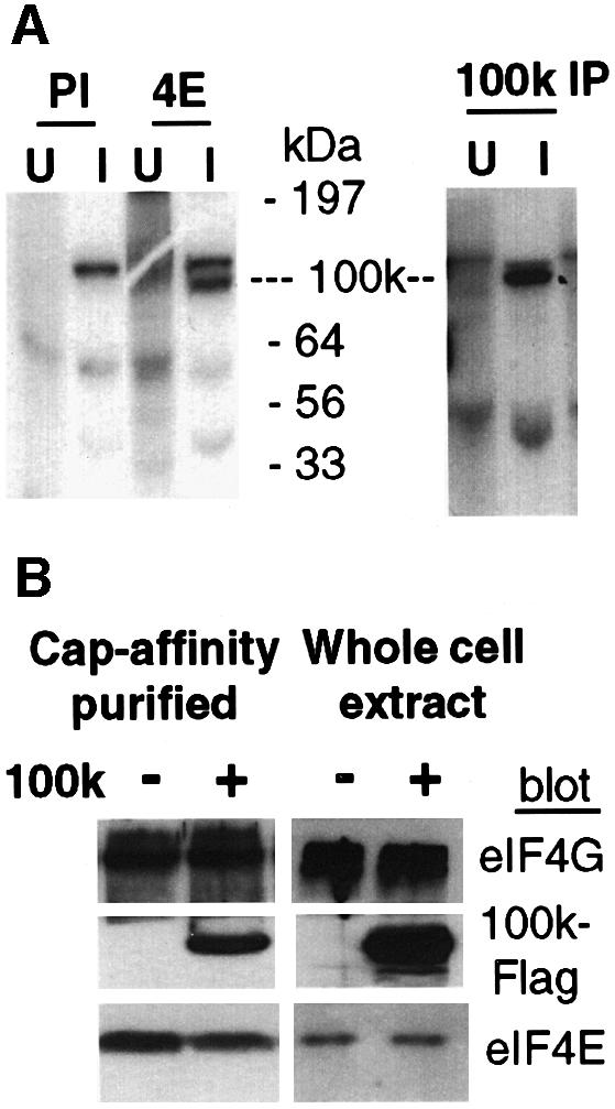
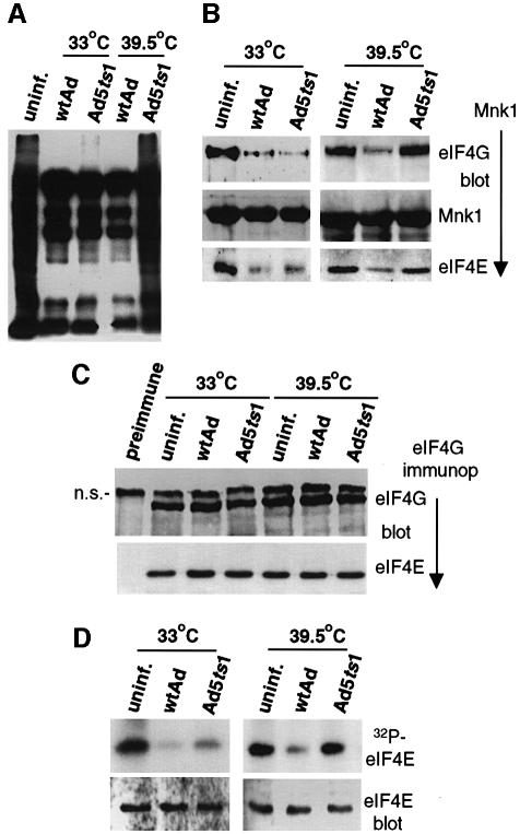
Translation of cellular mRNAs involves formation of a cap-binding translation initiation complex known as eIF4F, containing phosphorylated cap-binding protein eIF4E, eIF4E kinase Mnk1, eIF4A, poly(A)-binding protein and eIF4G. Adenovirus is shown to prevent cellular translation by displacing Mnk1 from eIF4F, thereby blocking phosphorylation of eIF4E. Over expression of an eIF4E mutant that cannot be phosphorylated by Mnk1 impairs translation of cellular but not viral late mRNAs. Adenovirus 100k protein is shown to bind the C-terminus of eIF4G in vivo and in vitro, the same region bound by Mnk1. In vivo, 100k protein displaces Mnk1 from eIF4G during adenovirus infection, or in transfected cells. Purified 100k protein also evicts Mnk1 from isolated eIF4F complexes in vitro. A mutant adenovirus with a temperature-sensitive 100k protein that cannot inhibit cellular protein synthesis at restrictive temperature no longer blocks Mnk1 binding to eIF4G, or phosphorylation of eIF4E. We describe a mechanism whereby adenovirus selectively inhibits the translation of cellular but not viral mRNAs by displacement of Mnk1 from eIF4G and inhibition of eIF4E phosphorylation.


Figures

None
Fig. 1. Ad does not impair Mnk1 kinase activity. 293 cells were transfected with plasmids expressing GST–Mnk1 or GST–T2A2, a kinase-deficient mutant of Mnk1 (Waskiewicz et al., 1997). Cells were either uninfected (uninf.), treated with 30 ng/ml EGF for 15 min (Feigenblum et al., 1998), or infected with wtAd for 36 h (late Ad inf.). GST–Mnk1 or GST–T2A2 was recovered from equal amounts of cell lysates by glutathione–Sepharose chromatography and incubated with 1 µg of purified recombinant eIF4E and [γ-32P]ATP, resolved by SDS–PAGE and autoradiographed (top panel), or subjected to immunoblot analysis with anti-GST antibodies (middle panel). eIF4E levels are shown by immunoblot (bottom panel). Quantitation was performed by digital densitometry.
None
Fig. 2. Immunoblot analysis of eIF4F complexes during Ad late infection. 293 cells were uninfected (uninf.) or infected for 36 h with wtAd (late Ad inf.). Equal amounts of protein lysates (10 µg) were resolved by SDS–10%PAGE and immunoblotted with specific antisera as described in Materials and methods. Antisera were: polyclonal anti-eIF4GI, polyclonal anti-GST (for Mnk1), monoclonal anti-eIF4A, polyclonal anti-eIF4E and monoclonal anti-PABP.
None
Fig. 3. Composition of eIF4F–Mnk1 complexes in Ad late infected cells. 293 cells were transfected with plasmids expressing GST or GST–Mnk1 proteins. At 18 h post-transfection, cells were uninfected or infected with wtAd and harvested at 12, 24 or 36 h post-infection (p.i.). (A) Cells were labeled with [35S]methionine and equal amounts of protein resolved by SDS–15%PAGE and fluorographed. (B) GST fusion proteins were recovered from equal amounts of cell lysates by glutathione–Sepharose chromatography, and immunoblot analysis of associated proteins performed using antisera to eIF4GI, GST (for Mnk1) and eIF4E. (C) Endogenous eIF4GI was immunoprecipitated (eIF4G immunop.) from equal amounts of cell lysates and associated proteins resolved by SDS–10%PAGE. Proteins were detected by immunoblot analysis using specific antisera as above. (D) Equal numbers of uninfected cells and cells infected for 12 h (early Ad infection) or 36 h (late Ad infection) were labeled with 200 µCi/ml of 32PO4 in phosphate-free medium for 2 h. eIF4E was recovered from equal amounts of cell extracts by m7GTP–Sepharose affinity chromatography, equal fractions resolved by SDS–15%PAGE and autoradiographed (upper panel), or transferred to Immobilon-P (Millipore) and detected by immunoblot analysis with specific eIF4E antisera (bottom panel). Quantitation was performed by digital densitometry.
None
Fig. 4. Effect of eIF4E phosphorylation on translation of model Ad late and cellular mRNAs. (A) 293T cells were cotransfected with plasmids expressing β-galactosidase mRNAs containing either the Ad late tripartite leader 5′UTR (3LDR), or an eIF4F-dependent 5′UTR (CR3), and plasmids expressing HA-tagged wt eIF4E, Ser209→Ala eIF4E or Ser209→Ala/Thr210→Ala eIF4E. At 36 h post-transfection, equal amounts of cell lysates were resolved by SDS–12%PAGE and immunoblotted using specific antisera for β-galactosidase protein (β-gal), HA-eIF4E and eIF4A (for protein levels). (B) 293 cells were transfected with plasmids expressing GST, GST–Mnk1 or the GST–T2A2 Mnk1 mutant. At 18 h post-transfection, cells were infected with wtAd for 36 h, or were uninfected, then labeled with [35S]methionine, or (C) labeled with 32PO4. Samples were analyzed as in the legend to Figure 3. Mnk proteins were detected by immunoblot analysis with antisera to GST. eIF4E phosphorylation and abundance were examined as in the legend to Figure 3.
None
Fig. 5. Association of Ad L4-100k protein with eIF4F complexes. 293 cells were uninfected (U) or infected (I) with wtAd. (A) Cells were labeled with 50 µCi/ml of [35S]methionine for 4 h at 24 h post-infection, lysates were prepared and equal amounts of protein were immunoprecipitated with preimmune sera (PI) or with antibodies specific for eIF4E (4E) or Ad5-100k protein (100k IP), followed by SDS–15%PAGE and autoradiography. (B) 293 cells were either mock transfected or transfected with a cDNA clone expressing Flag epitope tagged 100k protein. Equal amounts of lysates were prepared and eIF4F complexes were purified by cap-affinity chromatography (left panel), or directly resolved by SDS–10%PAGE (right panel). Proteins were detected by immunoblot analysis using antisera as in the legend to Figure 2.
None
Fig. 6. Characterization of 100k protein–eIF4F complexes in the absence of virus infection. (A) 293 cells were transfected with plasmids expressing GST, GST–Mnk1, GST–T2A2 protein or Flag-100k protein. At 36 h post-transfection, lysates were prepared from equal numbers of cells, GST fusion proteins were recovered by glutathione–Sepharose chromatography, resolved by SDS–10%PAGE, and associated proteins detected by immunoblot with antisera to endogenous eIF4GI, GST (for GST, GST–Mnk1, GST–T2A2) or endogenous eIF4E. (B) 293 cells were transfected with plasmids expressing Flag or Flag-100k protein, endogenous eIF4GI was immunoprecipitated with specific antisera (IP eIF4G), or preimmune sera (preimm). Immunoblotting was performed using antisera described in the legend to Figure 2. (C and D) Cells were transfected with plasmids expressing Flag-100k protein and full-length HA-eIF4GI (F4G, lacking the PABP binding site amino acids 157–1560); N-terminal fragment (N4G, 157–613), middle fragment (M4G, 565–1045) or C-terminal fragment (C4G, 1045–1560). (C) Flag-100k protein was immunoprecipitated (IP Flag-100k) with anti-Flag antibodies, proteins were resolved and immunoblotted for interaction with HA-eIF4G using anti-HA antibodies. Flag-100k proteins were detected using anti-Flag antibodies (bottom panel). (D) HA-eIF4G proteins were immunoprecipitated (IP HA-eIF4G) using anti-HA antibodies, proteins were resolved and immunoblotted as above.
None
Fig. 7. 100k protein binds the C-terminus of eIF4G and evicts Mnk1 protein from eIF4F complexes in vitro. (A) Soluble recombinant proteins were purified from bacteria corresponding to full-length GST–100k, GST–Mnk1 and fragments of eIF4G for the N-terminus (N4G), middle (M4G) and C-terminus (C4G). It was not possible to isolate sufficient amounts of full-length eIF4G due to degradation. GST was removed from eIF4G by thrombin digestion. Equal amounts of purified eIF4G proteins (2 µg) and GST–100k or GST–Mnk1 proteins (0.3 µg) were incubated in vitro, GST fusion proteins were recovered by glutathione–Sepharose chromatography and bound proteins identified by SDS–10%PAGE and immunoblot analysis with specific antibodies to the eIF4G fragments, or to GST (for Mnk1 or 100k). (B) 293 cells were transfected with plasmids expressing GST or GST–Mnk1, extracts were prepared and equal amounts of eIF4F isolated by immuno precipitation of eIF4G (eIF4G IP). Equal amounts (2 µg) of purified recombinant GST–100k or GST fusion proteins were incubated in vitro with eIF4F complexes, and associated proteins detected by immunoblot analysis as shown. Equal amounts of whole cell lysates (‘lysate’) were resolved in lanes 4 and 5 to establish protein levels.
None
Fig. 8. The ts-1 100k protein mutant is translationally inactive. 293 cells were transfected with a GST–Mnkl expression plasmid followed 18 h later by infection with wtAd or Ad5ts1. Cells were maintained at non-restrictive temperature (33°C) for 49 h, or restrictive temperature (39.5°C) for 29 h of infection. (A) Cells were labeled with [35S]methionine for 60 min, lysates prepared and equal amounts of protein resolved by SDS–10%PAGE and fluorographed. (B) Mnk1 was recovered from equal amounts of cell lysates by glutathione–Sepharose chromatography, or (C) eIF4G was immunoprecipitated, proteins were resolved by SDS–10%PAGE and detected by immunoblotting with specific antisera as shown. (D) Cells were labeled in vivo with [32P]orthophosphate at 33 or 39.5°C, eIF4E was recovered by m7GTP chromatography from equal amounts of lysate, resolved by SDS–15%PAGE and autoradiographed (upper panel), or subjected to immunoblot analysis with specific antisera as described in the legend to Figure 3 (lower panel). The protein migrating immediately above eIF4G is non-specific and also reacts with this preimmune serum.

Similar articles

Cited by

References

    1. Adam S.A. and Dreyfuss,G. (1987) Adenovirus proteins associated with mRNA and hnRNA in infected Hela cells. J. Virol., 61, 3276–3283. - PMC - PubMed
    1. Bonneau A.M. and Sonenberg,N. (1987) Involvement of the 24kd cap-binding protein in regulation of protein synthesis in mitosis. J. Biol. Chem., 262, 11134–11139. - PubMed
    1. Dolph P.J., Racaniello,V., Villamarin,A., Palladino,F. and Schneider,R.J. (1988) The adenovirus tripartite leader eliminates the requirement for cap binding protein during translation initiation. J. Virol., 62, 2059–2066. - PMC - PubMed
    1. Dolph P.J., Huang,J. and Schneider,R.J. (1990) Translation by the adenovirus tripartite leader: elements which determine independence from cap-binding protein complex. J. Virol., 64, 2669–2677. - PMC - PubMed
    1. Duncan R.F. (1996) Translational control during heat shock. In Hershey,J.W.B., Mathews,M.B. and Sonenberg,N. (eds), Translational Control During Heat Shock. Cold Spring Harbor Laboratory Press, Cold Spring Harbor, NY, pp. 271–294.

Publication types

MeSH terms

Substances

LinkOut - more resources