. 2000 Jul 3;19(13):3485-95.
doi: 10.1093/emboj/19.13.3485.

A geminivirus replication protein interacts with the retinoblastoma protein through a novel domain to determine symptoms and tissue specificity of infection in plants

Affiliations

A geminivirus replication protein interacts with the retinoblastoma protein through a novel domain to determine symptoms and tissue specificity of infection in plants

L J Kong et al. EMBO J. .

Abstract

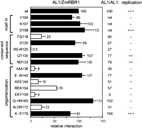
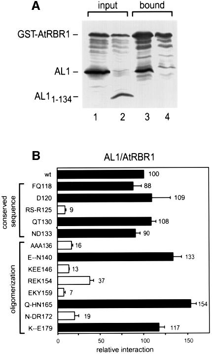
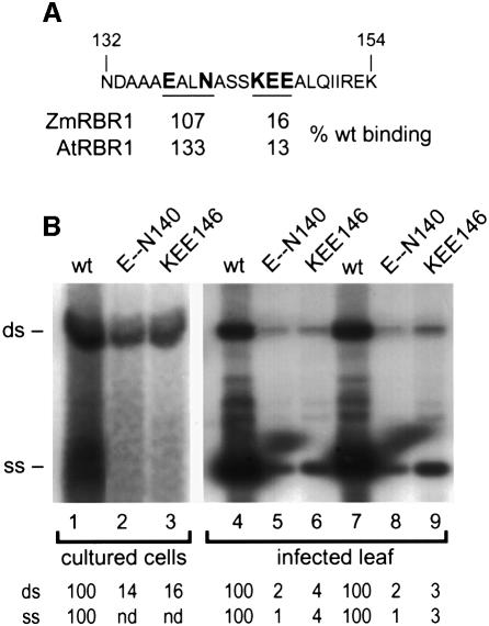
Geminiviruses replicate in nuclei of mature plant cells after inducing the accumulation of host DNA replication machinery. Earlier studies showed that the viral replication factor, AL1, is sufficient for host induction and interacts with the cell cycle regulator, retinoblastoma (pRb). Unlike other DNA virus proteins, AL1 does not contain the pRb binding consensus, LXCXE, and interacts with plant pRb homo logues (pRBR) through a novel amino acid sequence. We mapped the pRBR binding domain of AL1 between amino acids 101 and 180 and identified two mutants that are differentially impacted for AL1-pRBR interactions. Plants infected with the E-N140 mutant, which is wild-type for pRBR binding, developed wild-type symptoms and accumulated viral DNA and AL1 protein in epidermal, mesophyll and vascular cells of mature leaves. Plants inoculated with the KEE146 mutant, which retains 16% pRBR binding activity, only developed chlorosis along the veins, and viral DNA, AL1 protein and the host DNA synthesis factor, proliferating cell nuclear antigen, were localized to vascular tissue. These results established the importance of AL1-pRBR interactions during geminivirus infection of plants.


Figures

None
Fig. 1. AL1 and large T-antigen interact with ZmRBR1 differently. (A) Diagram of the maize pRb homologue ZmRBR1 showing the pocket domain with the A and B boxes. Arrows mark the N-terminal truncations at positions 214 and 290 and the C653F mutation. (B) Mean β-galactosidase specific activities (1 unit = 1.0 mmol product/min/mg protein at pH 7.3 at 37°C) from two-hybrid assays containing the indicated GAL4 DBD–ZmRBR1 fusions and GAL4 AD fusions for full-length TGMV AL1 or SV40 large T-antigen (amino acids 87–708) are given. Two standard errors are given for each value. Relative activities are in parentheses.
None
Fig. 2. Mapping the pRBR binding domain of TGMV AL1. (A) Diagram of the AL1 protein showing the positions of the three DNA cleavage motifs (solid boxes), two predicted pairs of α-helices (hatched ovals) and the ATP binding site (hatched box). The DNA binding and cleavage/ligation domains are indicated by solid lines and the oligomerization domain is shown as a dashed line. Solid lines below the diagram mark the sizes of truncated AL1 proteins, which are designated by their N- and C-terminal amino acids. The boxed region indicates the limits of the pRBR binding domain. (B) Total protein extracts from insect cells co-expressing GST or GST–ZmRBR1 with different AL1 proteins (top) were incubated with glutathione–Sepharose, washed and eluted. Input (lanes 1–6) and bound (lanes 7–12) proteins were resolved by SDS–PAGE and analyzed by immunoblotting. The top panels were visualized using an anti-AL1 antibody while the bottom panels were visualized using an anti-GST antibody. The extracts in lanes 1 and 7 contained GST and full-length AL11–352. The extracts in lanes 2–6 and 8–12 contained GST–ZmRBR1 and full-length AL11–352 (lanes 2 and 8), AL1181–352 (lanes 3 and 9), AL11–213 (lanes 4 and 10), AL11–180 (lanes 5 and 11) or AL11–119 (lanes 6 and 12). (C) Input (lanes 1–3) and bound (lanes 4–6) fractions are shown for interactions between GST–ZmRBR1 and the C-terminal truncations (top) corresponding to AL11–180 (lanes 1 and 4), AL11–168 (lanes 2 and 5) and AL11–158 (lanes 3 and 6). (D) Input (lanes 1–3) and bound (lanes 4–6) fractions are shown for interactions between GST–ZmRBR1 and the N-terminal truncations (top) corresponding to AL1101–352 (lanes 1 and 4), AL1110–352 (lanes 2 and 5) and AL1119–352 (lanes 3 and 6). In (C) and (D), the blots were probed with both anti-AL1 and anti-GST antibodies.
None
Fig. 3. Site-directed mutations in the pRBR binding domain of TGMV AL1. The AL1 sequence between amino acids 100 and 180 is shown, and the locations of motif III, a conserved sequence and the oligomerization domain are indicated by the solid and dotted lines above the sequence. The predicted α-helices 3 and 4 are also marked. The boxed region indicates the positions of the site-directed substitutions. Mutations (on the left) are designated by the wild-type sequence and the position of the last altered amino acid. Dashes indicate amino acids that were not changed.
None
Fig. 4. Mutations in the pRBR binding domain of AL1 impair interactions with ZmRBR1. A ZmRBR1 expression cassette corresponding to Zm214C fused to the GAL4 DBD was co-transformed into yeast with cassettes for either wild-type or mutant AL1 fused to the GAL4 AD (on the left). Interactions between ZmRBR1 and the AL1 proteins were assayed by measuring β-galactosidase activity in total protein extracts and normalized to wild type (100). The open bars indicate mutants impaired for ZmRBR1 binding, whereas the filled bars mark mutants with activity similar to or greater than wild-type AL1. The error bars correspond to two standard errors. The locations of motif III, the conserved sequence and the oligomerization domain are shown on the left. The effects of the mutations on AL1 oligomerization activity and TGMV replication in transient assays are indicated on the right (Orozco et al., 2000).
None
Fig. 5. AL1 interacts with AtRBR1. (A) Total protein extracts from insect cells co-expressing GST–AtRBR1 with either full-length AL11–352 (lanes 1 and 3) or AL11–134 (lanes 2 and 4) were incubated with glutathione–Sepharose, washed and eluted. Input (lanes 1 and 2) and bound (lanes 3 and 4) proteins were resolved by SDS–PAGE and analyzed by immunoblotting with anti-AL1 and anti-GST antibodies. (B) An AtRBR1 expression cassette corresponding to At319C fused to the GAL4 DBD was cotransformed into yeast with cassettes for either wild-type or mutant AL1 fused to the GAL4 AD (on the left) and analyzed as described in Figure 4. The locations of the conserved sequence and the oligomerization domain are indicated on the left.
None
Fig. 6. Two TGMV A mutants altered in the AL1 pRBR binding domain replicate to similar levels. (A) The sequence between AL1 amino acids 132 and 154 is shown, with the mutated amino acids in E--N140 and KEE146 indicated by bold face type. The relative binding activities with ZmRBR1 and AtRBR1 are given for the two mutants. (B) In lanes 1–3, tobacco protoplasts were transfected with TGMV A replicons with either wild-type (lane 1) or mutant AL1 ORFs corresponding to E--N140 (lane 2) or KEE146 (lane 3). Total DNA was isolated from 7 × 10⁶ cells 72 h post-transfection and analyzed on DNA gel blots. In lanes 4–9, N.benthamiana plants were bombarded with DNAs corresponding to TGMV A and B replicons. The AL1 ORFs of the A components were either wild type (lanes 4 and 7) or carried the E--N140 (lanes 5 and 8) or KEE146 mutation (lanes 6 and 9). Total DNA (2.5 µg/lane) was isolated from systemically infected leaves from two plants for each construct at 18 days post-infection and analyzed on DNA gel blots. TGMV DNA was detected using a radiolabeled probe specific for the A component. The positions of double- (ds) and single- (ss) stranded forms of TGMV A are marked on the left. The relative accumulations of the DNA forms are given at the bottom of each lane with wild type set at 100. In lanes 2 and 3, single-stranded DNA was not detected (nd).
None
Fig. 7. A pRBR binding mutant displays altered symptoms and accumulation patterns in plants. Nicotiana benthamiana plants infected with the pRBR binding mutant KEE146 (AE), the control mutant E-N140 (F, G and H) or wild-type TGMV (I, J and K) were analyzed at 18 days post-infection. (A), (F) and (I) show symptomatic plants. In (B), (G) and (J), viral DNA was detected in mature leaves by in situ hybridization with a TGMV B probe labeled with digoxigenin and visualized using an anti-digoxigenin antibody conjugated to alkaline phosphatase. In (D), (H) and (K), TGMV AL1 protein was detected using an anti-AL1 antibody and peroxidase detection. In (E), the host DNA synthesis factor, PCNA, was detected using an anti-human PCNA antibody and peroxidase detection. Total DNA was visualized in (C) by DAPI staining, with the same section depicted in (B) and (C). Arrows in (B), (D) and (E) show nuclei infected with the KEE146 mutant. All sections were analyzed at 400× magnification.

Similar articles

Cited by

References

    1. Ach R.A., Durfee,T., Miller,A.B., Taranto,P., Hanley-Bowdoin,L., Zambriski,P.C. and Gruissem,W. (1997a) RRB1 and RRB2 encode maize retinoblastoma-related proteins that interact with a plant D-type cyclin and geminivirus replication protein. Mol. Cell. Biol., 17, 5077–5086. - PMC - PubMed
    1. Ach R.A., Taranto,P. and Gruissem,W. (1997b) A conserved family of WD-40 proteins binds to the retinoblastoma protein in both plants and animals. Plant Cell, 9, 1595–1606. - PMC - PubMed
    1. Aronson M.N., Meyer,A.D., Gyorgyey,J., Katul,L., Vetten,H.J., Gronenborn,B. and Teimchenko,T. (2000) Clink, a nanovirus-encoded protein, binds both pRB and SKP1. J. Virol., 74, 2967–2972. - PMC - PubMed
    1. Bass H., Nagar,S., Hanley-Bowdoin,L. and Robertson,D. (2000) Chromosome condensation induced by geminivirus infection of mature plant cells. J. Cell Sci., 113, 1149–1160. - PubMed
    1. Chellappan S., Kraus,V.B., Kroger,B., Munger,K., Howley,P.M., Phelps,W.C. and Nevins,J.R. (1992) Adenovirus E1A, simian virus 40 tumor antigen and human papillomavirus E7 protein share the capacity to disrupt the interaction between transcription factor E2F and the retinoblastoma gene product. Proc. Natl Acad. Sci. USA, 89, 4549–4553. - PMC - PubMed

Publication types

LinkOut - more resources