. 2000 Jul 3;192(1):1-10.
doi: 10.1084/jem.192.1.1.

The semiconserved head structure of Plasmodium falciparum erythrocyte membrane protein 1 mediates binding to multiple independent host receptors

Affiliations

The semiconserved head structure of Plasmodium falciparum erythrocyte membrane protein 1 mediates binding to multiple independent host receptors

Q Chen et al. J Exp Med. .

Abstract

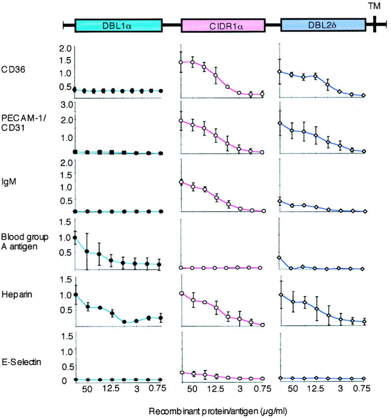
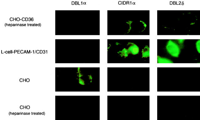
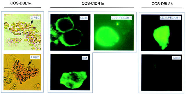
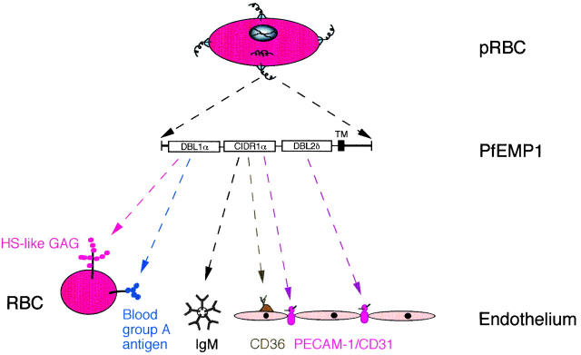
Erythrocytes infected with mature forms of Plasmodium falciparum do not circulate but are withdrawn from the peripheral circulation; they are bound to the endothelial lining and to uninfected erythrocytes in the microvasculature. Blockage of the blood flow, hampered oxygen delivery, and severe malaria may follow if binding is excessive. The NH(2)-terminal head structure (Duffy binding-like domain 1 [DBL1alpha]-cysteine-rich interdomain region [CIDR1alpha]) of a single species of P. falciparum erythrocyte membrane protein 1 (PfEMP1) is here shown to mediate adherence to multiple host receptors including platelet-endothelial cell adhesion molecule 1 (PECAM-1)/CD31, the blood group A antigen, normal nonimmune immunoglobulin M, three virulence-associated receptor proteins, a heparan sulfate-like glucosaminoglycan, and CD36. DBL2delta was found to mediate additional binding to PECAM-1/CD31. The exceptional binding activity of the PfEMP1 head structure and its relatively conserved nature argues that it holds an important role in erythrocyte sequestration and therefore in the virulence of the malaria parasite.


Figures

Figure 1
Figure 1
The binding of fluorescence-labeled soluble receptors to the surface of erythrocytes infected with FCR3S1.2. Fluorescence-labeled recombinant human CD36, ICAM-1, or PECAM-1/CD31 was directly incubated with the infected cells. The binding of human nonimmune IgM was detected by incubating the pRBCs with FITC-labeled rabbit anti–human Ig, whereas that of the blood group antigen A was by incubating the infected cells with an A trisaccharide coupled to biotinylated BSA and FITC-avidin. Infected (arrows) and uninfected RBCs are shown with phase–contrast (PC) and in parallel with UV light (UV). The binding shown was at a receptor concentration of 100 μg/ml, and the parasites were counterstained with ethidium bromide. The fluorescence rates were 65–70% (CD36), 0–5% (ICAM-1), 70–80% (PECAM-1/CD31), 75–85% (IgM), and 60–70% (blood group A). For further details, see Materials and Methods.
Figure 2
Figure 2
The interaction of the recombinant PfEMP1 domains (DBL1α, CIDR1α, and DBL2δ) as GST fusion proteins with different receptors (CD36, PECAM-1/CD31, blood group antigen A, heparin, and E-selectin) or receptor proteins (IgM) bound to solid phase as measured by ELISA. The domain-like primary structure of FCR3S1.2var1-PfEMP1 is shown at the top of the figure (colored boxes). Each receptor studied (left) for interaction with a GST fusion protein is shown as a separate curve (DBL1α, blue •; CIDR1α, pink ○; DBL2δ, violet ⋄). The ranges of absorption values are shown on the y-axis and the concentrations (in μg/ml) on the x-axis. The results are the mean of at least three separate experiments ± 2 SD. For further details, see Materials and Methods.
Figure 3
Figure 3
The interaction of the recombinant PfEMP1 domains (DBL1α, CIDR1α, and DBL2δ) as GST fusion proteins with receptors expressed on normal or transfected cells (some of the cells were treated with heparinase to remove HS from the cell surface) visualized by a fluorescence-labeled anti-GST antibody. The results are those of at least three separate experiments. 95% untreated and 5% treated CHO cells (CHO and CHO-CD36) bind DBL1α, respectively. More than 80% untreated CHO-CD36 cells bind CIDR1α, whereas <30% CHO-CD36 bind DBL2δ. For further details, see Materials and Methods.
Figure 4
Figure 4
The binding of fluorescence-labeled receptors, receptor proteins, or erythrocytes to COS-7 cells transiently transfected with the different PfEMP1 domains (DBL1α, CIDR1α, and DBL2δ) of FCR3S1.2var1. The transfected cell type used is shown at the top of the figure. The fluorescence-labeled protein or erythrocyte used is shown in the right hand corner of each photograph. The results are those of at least three separate experiments. For further details, see Materials and Methods.
Figure 5
Figure 5
Schematic summary of the different binding activities of the DBL1α, CIDR1α, and DBL2δ domains of PfEMP1 encoded by FCR3S1.2var1. P. falciparum expresses PfEMP1 molecules (here shown corkscrew-like) on a knob-like structure at the infected erythrocyte surface. The DBL1α domain participates in rosetting through binding to an HS-like GAG and to the blood group A antigen. The CIDR1α domain binds to CD36 and to members of the immunoglobulin superfamily, including IgM and PECAM-1/CD31, whereas the DBL2δ domain binds mainly to PECAM-1/CD31.

References

    1. Miller L.H., Good M.F., Milon G. Malaria pathogenesis. Science. 1994;264:1878–1883. - PubMed
    1. Barnwell J.W., Asch A.S., Nachman R.L., Yamaya M., Aikawa M., Ingravallo P. A human 88-kD membrane glucoprotein (CD36) functions in vitro as a receptor for a cytoadherence ligand on Plasmodium falciparum–infected erythrocytes. J. Clin. Invest. 1989;84:77–87. - PMC - PubMed
    1. Berendt A.R., Simmons D.L., Tansey J., Newbold C.I., Marsh K. Intercellular adhesion molecule-1 is an endothelial cell adhesion receptor for Plasmodium falciparum . Nature. 1989;341:57–59. - PubMed
    1. Robert C., Pouvelle B., Meyer P., Muanza K., Fujioka H., Aikawa M., Scherf A., Gysin J. Chondroitin-4-sulfate (proteoglycan), a receptor for Plasmodium falciparum-erythrocyte adhesion. Res. Immunol. 1995;146:383–393. - PubMed
    1. Rogerson S.J., Chaiyaroj C.S., Ng K., Reeder J.C., Brown G.V. Chondroitin sulfate A is a cell surface receptor for Plasmodium falciparum–infected erythrocytes. J. Exp. Med. 1995;182:15–20. - PMC - PubMed

Publication types

MeSH terms

Substances