. 2000 Jul 3;192(1):63-75.
doi: 10.1084/jem.192.1.63.

HIV-specific CD8(+) T cells produce antiviral cytokines but are impaired in cytolytic function

Affiliations

HIV-specific CD8(+) T cells produce antiviral cytokines but are impaired in cytolytic function

V Appay et al. J Exp Med. .

Abstract

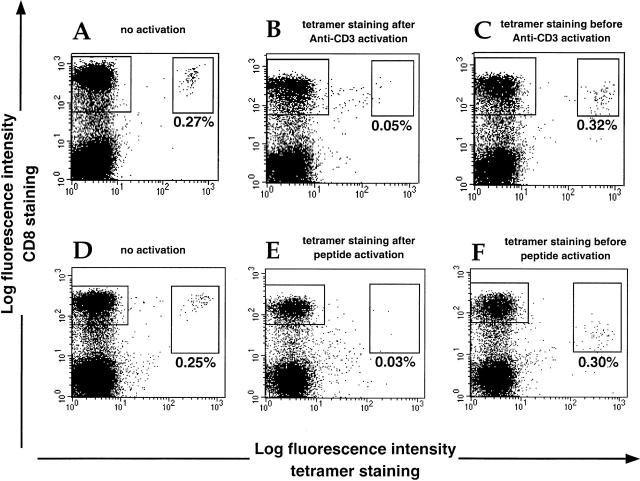
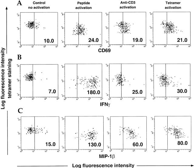
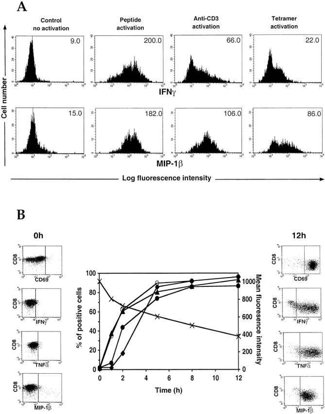
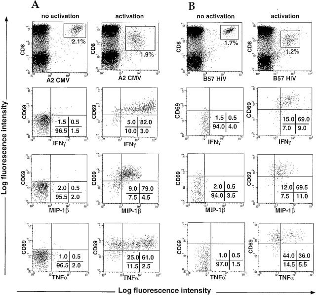
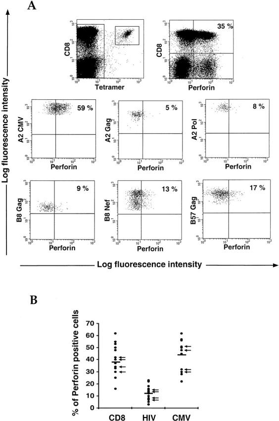
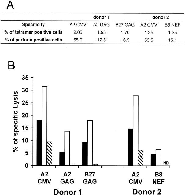
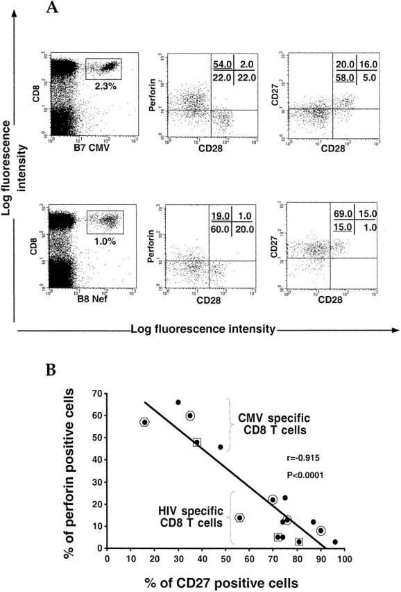
The use of peptide-human histocompatibility leukocyte antigen (HLA) class I tetrameric complexes to identify antigen-specific CD8(+) T cells has provided a major development in our understanding of their role in controlling viral infections. However, questions remain about the exact function of these cells, particularly in HIV infection. Virus-specific cytotoxic T lymphocytes exert much of their activity by secreting soluble factors such as cytokines and chemokines. We describe here a method that combines the use of tetramers and intracellular staining to examine the functional heterogeneity of antigen-specific CD8(+) T cells ex vivo. After stimulation by specific peptide antigen, secretion of interferon (IFN)-gamma, tumor necrosis factor (TNF)-alpha, macrophage inflammatory protein (MIP)-1beta, and perforin is analyzed by FACS((R)) within the tetramer-positive population in peripheral blood. Using this method, we have assessed the functional phenotype of HIV-specific CD8(+) T cells compared with cytomegalovirus (CMV)-specific CD8(+) T cells in HIV chronic infection. We show that the majority of circulating CD8(+) T cells specific for CMV and HIV antigens are functionally active with regards to the secretion of antiviral cytokines in response to antigen, although a subset of tetramer-staining cells was identified that secretes IFN-gamma and MIP-1beta but not TNF-alpha. However, a striking finding is that HIV-specific CD8(+) T cells express significantly lower levels of perforin than CMV-specific CD8(+) T cells. This lack of perforin is linked with persistent CD27 expression on HIV-specific cells, suggesting impaired maturation, and specific lysis ex vivo is lower for HIV-specific compared with CMV-specific cells from the same donor. Thus, HIV-specific CD8(+) T cells are impaired in cytolytic activity.


Figures

Figure 1
Figure 1
Tetramer staining and cell activation. PBMCs from a CMV-positive B7 donor were incubated for 12 h with brefeldin A in the presence of PBS (no activation control) (A and D), OKT3 (100 ng/ml) (B and C), or specific CMV B7 peptide (10 μM) (E and F). Tetramer staining was carried out either after overnight incubation (A, B, D, and E) or before addition of the activators (C and F). Percentages for tetramer-staining cells are shown. Cells were fixed and analyzed by flow cytometry after staining for CD8 surface molecules.
Figure 2
Figure 2
Comparison among peptide, OKT3, and tetramer activation. PBMCs from a CMV-positive B7 donor were incubated for 12 h with brefeldin A in the presence of PBS (no activation control), specific CMV B7 peptide (10 μM), OKT3 (100 ng/ml), or CMV B7 tetramers. Tetramer staining was carried out after overnight incubation for the no activation control or before addition of the activators for the “activated” cells. Cells were stained for CD69 (A), IFN-γ (B), and MIP-1β (C) and analyzed by flow cytometry. Data show cells gated on the CMV B7 tetramer-positive population. Mean fluorescence intensity is shown for each condition.
Figure 3
Figure 3
Intracellular staining in CTL clones. (A) A CMV-specific B7 clone was incubated for 12 h with brefeldin A in the presence of PBS (no activation control), specific antigen-pulsed APCs, OKT3 (coated onto the experimental wells), or CMV B7 tetramers. Intracellular staining for IFN-γ and MIP-1β was carried out, and the cells were analyzed by flow cytometry. Mean fluorescence intensity is shown for each condition. (B) An HIV-specific Cw4-restricted CTL clone was incubated for various times in the presence of brefeldin A and specific antigen-pulsed APCs. Cells were stained and analyzed by flow cytometry. CD8 staining (x) is expressed in mean fluorescence intensity, and CD69 (○), IFN-γ (•), TNF-α (▴), and MIP-1β (♦) stainings are expressed in percentage of positive cells.
Figure 4
Figure 4
Intracellular staining for IFN-γ, MIP-1β, and TNF-α in HIV-specific or CMV-specific CD8+ T cells. PBMCs from HIV-infected patients were incubated for 6 h with brefeldin A in the presence of PBS (no activation) or specific peptides (10 μM) (activation). Intracellular staining for IFN-γ, MIP-1β, and TNF-α was carried out, and the cells were analyzed by flow cytometry. Cytokine staining is shown on CMV (A) and HIV (B) specific CD8+ T cell populations gated using the tetramers (top). Percentages of cells present in quadrants are shown. Representative data are shown (see Table ).
Figure 5
Figure 5
Perforin staining in antigen-specific CD8+ T cells. (A) Perforin intracellular staining was carried out in a sample from an HIV-infected patient known to have both HIV-specific and CMV-specific CD8+ T cell populations. Cells were stained with tetramers and directly stained for perforin without cell activation. Both the total lymphocyte population (top row) and the antigen-specific CD8+ T cells (bottom rows) were gated and analyzed for perforin staining. The data are expressed as percentages of perforin-positive cells determined to be staining above the horizontal limit. Similar observations were obtained with other donors who also had both HIV-specific and CMV-specific CD8+ T cell populations. (B) Perforin intracellular staining was performed, as above, in samples known to have either an HIV-specific or a CMV-specific CD8 T cell population. Data are expressed in numbers of perforin-positive cells within the relevant cell populations. 18, 18, and 13 samples are displayed for whole, HIV-specific, and CMV-specific CD8+ T cell populations, respectively. Arrows indicate that samples come from HAART-treated donors.
Figure 6
Figure 6
Fresh cytotoxic assay with HIV-specific or CMV-specific CD8+ T cells from the same donors. (A) Cell frequency and percentage of HIV-specific or CMV-specific CD8+ T cells expressing perforin from each donor. (B) Specific lysis obtained with the relevant HIV-specific or CMV-specific populations. Freshly isolated PBMCs were incubated for 5 h with targets pulsed with specific peptide or no peptide at different E/T ratios: 100:1 (black bars), 200:1 (white bars), or 100:1 after partial depletion of the tetramer-positive cells (hatched bars).
Figure 7
Figure 7
Comparison between CMV-specific and HIV-specific CD8+ T cells for perforin and maturation markers. (A) Stainings for perforin, CD28, and CD27 were performed in CMV (B7 CMV) or HIV (B8 Nef) specific CD8+ T cell populations gated by means of specific tetramers. Percentages of cells present in quadrants are shown. Data are representative of 15 different stainings. (B) Inverse correlation between perforin and CD27 staining in HIV-specific and CMV-specific CD8+ T cells. Each dot represents perforin and CD27 staining for a single antigen-specific CD8+ T cell population. Framed dots (circle, square, or hexagon) show specific populations belonging to the same donor.

References

    1. Carmichael A., Jin X., Sissons P., Borysiewicz L. Quantitative analysis of the human immunodeficiency virus type 1 (HIV-1)–specific cytotoxic T lymphocyte (CTL) response at different stages of HIV-1 infectiondifferential CTL responses to HIV-1 and Epstein-Barr virus in late disease. J. Exp. Med. 1993;177:249–256. - PMC - PubMed
    1. Kalams S.A., Johnson R.P., Trocha A.K., Dynan M.J., Ngo S., D'Aquila R.T., Kurnick J.T., Walker B.D. Longitudinal analysis of T cell receptor (TCR) gene usage by human immunodeficiency virus 1 envelope–specific cytotoxic T lymphocyte clones reveals a limited TCR repertoire. J. Exp. Med. 1994;179:1261–1271. - PMC - PubMed
    1. Moss P.A.H., Rowland-Jones S.L., Frodsham P.M., McAdam S., Giangrande P., McMichael A.J., Bell J.I. Persistent high frequency of human immunodeficiency virus-specific cytotoxic T cells in peripheral blood of infected donors. Proc. Natl. Acad. Sci. USA. 1995;92:5773–5777. - PMC - PubMed
    1. Kern F., Surel I.P., Brock C., Freistedt B., Radtke H., Scheffold A., Blasczyk R., Reinke P., Schneider-Mergener J., Radbruch A. T-cell epitope mapping by flow cytometry. Nat. Med. 1998;4:975–978. - PubMed
    1. Lalvani A., Brookes R., Hambleton S., Britton W.J., Hill A.V., McMichael A.J. Rapid effector function in CD8+ memory T cells. J. Exp. Med. 1997;186:859–865. - PMC - PubMed

Publication types

MeSH terms