. 2000 Jul 3;192(1):117-22.
doi: 10.1084/jem.192.1.117.

Interferon gamma eliminates responding CD4 T cells during mycobacterial infection by inducing apoptosis of activated CD4 T cells

Affiliations

Interferon gamma eliminates responding CD4 T cells during mycobacterial infection by inducing apoptosis of activated CD4 T cells

D K Dalton et al. J Exp Med. .

Abstract

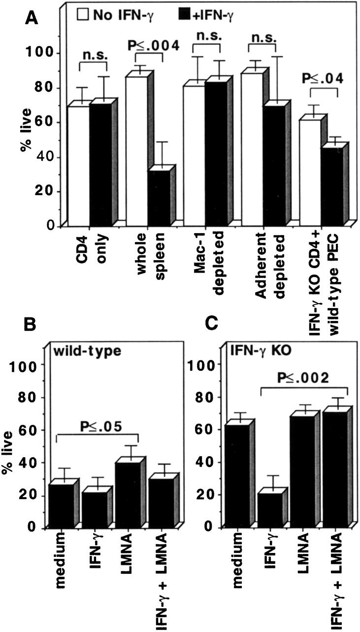
In Mycobacterium bovis Bacille Calmette-Guérin (BCG)-infected wild-type mice, there was a large expansion of an activated (CD44(hi)) splenic CD4 T cell population followed by a rapid contraction of this population to normal numbers. Contraction of the activated CD4 T cell population in wild-type mice was associated with increased apoptosis of activated CD4 T cells. In BCG-infected interferon (IFN)-gamma knockout (KO) mice, the activated CD4 T cell population did not undergo apoptosis. These mice accumulated large numbers of CD4(+)CD44(hi) T cells that were responsive to mycobacterial antigens. Addition of IFN-gamma to cultured splenocytes from BCG-infected IFN-gamma KO mice induced apoptosis of activated CD4 T cells. IFN-gamma-mediated apoptosis was abolished by depleting adherent cells or Mac-1(+) spleen cells or by inhibiting nitric oxide synthase. Thus, IFN-gamma is essential to a regulatory mechanism that eliminates activated CD4 T cells and maintains CD4 T cell homeostasis during an immune response.


Figures

Figure 1
Figure 1
Kinetics of expansion and contraction of activated CD4 T cells during BCG infection. (A) Typical FACS® histogram plots of CD44 expression on live CD4 T cells of uninfected mice or BCG-infected wild-type and IFN-γ KO mice at 4.5 wk of BCG infection. The CD44 marker was always set on uninfected mice as shown. The CD4 T cell population of IFN-γ KO mice is extremely skewed toward high levels of expression of CD44. (B) Total number of CD4+CD44hi T cells in the spleens of wild-type and IFN-γ KO mice during BCG infection. Cells were stained and analyzed by flow cytometry to measure CD4 and CD44 expression. Shown is the total number of CD4+CD44hi T cells, calculated as: percentage of live cells that are CD4+CD44hi × total number of live splenocytes. Numbers shown are average and SD of six to eight mice per time point, done in two to three experiments per time point. (C) Kinetics of expression of high levels of CD44 on splenic CD4 T cells during BCG infection. Numbers shown are percentage of live CD4 T cells with CD44hi. This is the average and SD of six to eight mice per group, done in replicate experiments.
Figure 2
Figure 2
Comparison of the proliferative response of enriched CD4 T cells to mycobacterial antigens. Shown is the Δ cpm: cpm of CD4 T cells cultured with APCs prepulsed with 10 μg/ml antigen − cpm of CD4 T cells incubated with APCs and no antigens.
Figure 3
Figure 3
Measurement of apoptosis of CD4+CD44hi T cells ex vivo during BCG infection. (A) Typical dot plots of annexin V and PI staining of gated CD4+CD44hi T cells in spleens of wild-type and IFN-γ KO mice at 4 wk of BCG infection. (B) Kinetics of apoptosis of gated CD4+CD44hi T cells from the spleens of BCG-infected wild-type and IFN-γ KO mice. A gate was set on CD4+CD44hi T cells, and the percentage of apoptotic CD4+CD44hi T cells was determined by annexin V and PI staining. Numbers shown are the percentages of cells that are in the early stages of apoptosis. Each point is the average and SD of at least six to eight mice done in two to three or more separate experiments. (C) Apoptosis of gated CD4+CD44hi T cells from the spleens of BCG-infected wild-type, lpr/lpr (on C57BL/6), and IFN-γ KO mice; all three groups were compared in the same experiment each time. Numbers shown are percentages of CD4+CD44hi T cells in early apoptosis. Each point is the average and SD of six mice done in two separate experiments.
Figure 4
Figure 4
Induction of apoptosis of CD4+CD44hi T cells in vitro by IFN-γ. Spleen cells from BCG-infected wild-type and IFN-γ KO mice, week 3 or 4 after infection, were cultured in medium with IL-2, with or without IFN-γ. A gate was set on CD4+CD44hi T cells, and the percentage of cells that were live, apoptotic, and dead at 65 h of culture was determined from the annexin V/PI dot plots. Each bar is the average and SD of six mice per group, done in two different experiments. Statistical significance (t test) between groups is shown.
Figure 5
Figure 5
Activated macrophages and NO were required for IFN-γ to induce apoptosis of activated CD4 T cells. (A) The cells indicated, from BCG-infected IFN-γ KO mice or IFN-γ KO CD4 T cells with wild-type PECs, all at week 3–4 of BCG infection, were cultured in the presence or absence of IFN-γ. The percentage of live CD4+CD44hi T cells at 65 h of culture was measured using the annexin/PI assay. Average and SD of three to nine mice done in multiple experiments. (B) Wild-type and (C) IFN-γ KO spleen cells, used at week 3–4 of BCG infection, were cultured in the indicated conditions. Percent live CD4+CD44hi cells at 65 h is shown. Statistical significance (t test) between groups is shown.

References

    1. Murali-Krishna K., Altman J.D., Suresh M., Sourdive D.J., Zajac A.J., Miller J.D., Slansky J., Ahmed R. Counting antigen-specific CD8 T cellsa reevaluation of bystander activation during viral infection. Immunity. 1998;8:177–187. - PubMed
    1. Van Parijs L., Abbas A.K. Homeostasis and self-tolerance in the immune systemturning lymphocytes off. Science. 1998;280:243–248. - PubMed
    1. Chambers C.A., Krummel M.F., Boitel B., Hurwitz A., Sullivan T.J., Fournier S., Cassell D., Brunner M., Allison J.P. The role of CTLA-4 in the regulation and initiation of T-cell responses. Immunol. Rev. 1996;153:27–46. - PubMed
    1. Van Parijs L., Peterson D.A., Abbas A.K. The Fas/Fas ligand pathway and Bcl-2 regulate T cell responses to model self and foreign antigens. Immunity. 1998;8:265–274. - PubMed
    1. Bachmann M.F., Waterhouse P., Speiser D.E., McKall-Faienza K., Mak T.W., Ohashi P.S. Normal responsiveness of CTLA-4-deficient anti-viral cytotoxic T cells. J. Immunol. 1998;160:95–100. - PubMed

Publication types

MeSH terms