. 2000 Jul 3;192(1):145-50.
doi: 10.1084/jem.192.1.145.

Immature dendritic cells acquire CD8(+) cytotoxic T lymphocyte priming capacity upon activation by T helper cell-independent or -dependent stimuli

Affiliations

Immature dendritic cells acquire CD8(+) cytotoxic T lymphocyte priming capacity upon activation by T helper cell-independent or -dependent stimuli

D H Schuurhuis et al. J Exp Med. .

Abstract

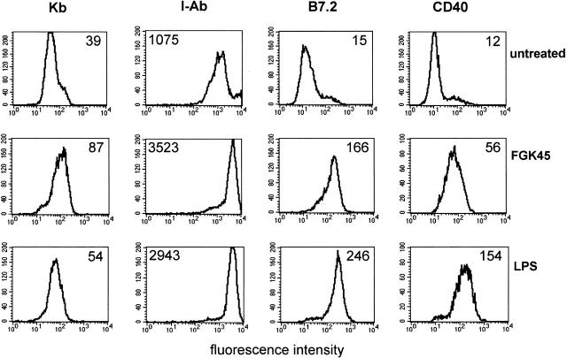
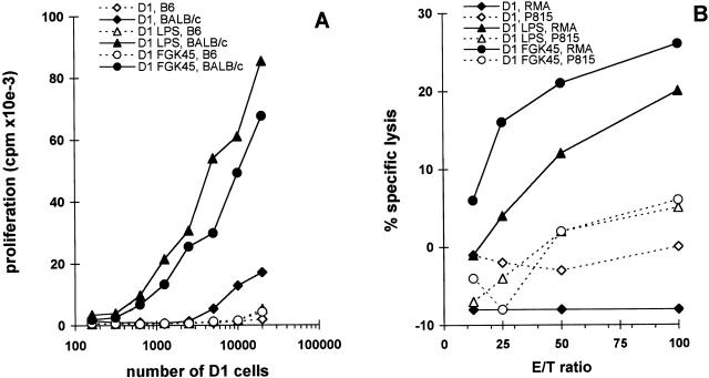
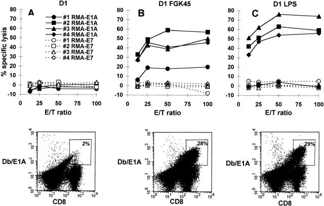
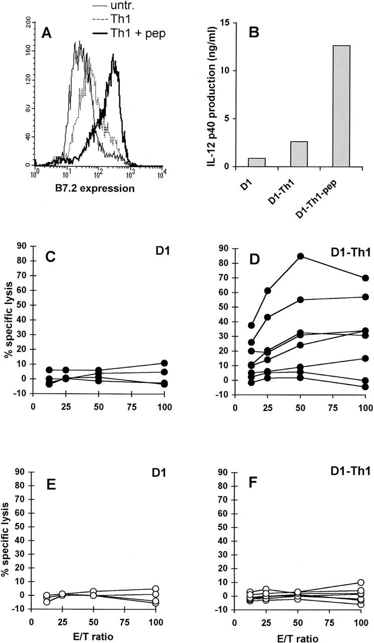
The well defined, immature murine dendritic cell (DC) line D1 was used to study the role of DC maturation in CTL induction in vitro and in vivo. Maturation of D1 cells, characterized by markedly increased expression of MHC and costimulatory molecules, was induced by incubation with lipopolysaccharide, agonistic CD40 antibody, or specific CD4(+) T helper (Th) cells. Activated, but not immature, D1 cells efficiently primed alloreactive T cell responses in vitro. Similarly, priming of CTL immunity in vivo in CD4-depleted mice was only observed if these mice were immunized with activated D1 cells. This study provides formal evidence that activation of DCs, induced by Th-independent as well as Th-dependent stimuli, is essential for efficient induction of CTL responses.


Figures

Figure 1
Figure 1
Both agonistic CD40 antibody and LPS treatment induce phenotypic maturation of D1 cells. D1 cells were treated with LPS or CD40-stimulating antibody FGK45 or left untreated (immature) for 48 h and stained with antibodies against the indicated markers. Data from one representative experiment out of three experiments performed are shown. Numbers indicate the median fluorescence intensity.
Figure 2
Figure 2
Efficient induction of primary allospecific responses in vitro by mature D1 cells. D1 cells (H-2b), treated with agonistic CD40 antibody FGK45 or LPS for 48 h, were used as stimulator cells in a proliferation assay with 105 BALB/c (H-2d) or control syngeneic B6 (H-2b) responder cells (A) or as stimulator cells in bulk cultures for the induction of allospecific CTLs (B). For CTL induction, 104 D1 cells were incubated with 3 × 10⁶ BALB/c spleen cells for 6 d. CTL activity was measured in a cytotoxicity assay using targets of H-2b haplotype (RMA) or H-2d haplotype (P815). Values represent means of triplicates from one representative experiment out of three performed.
Figure 3
Figure 3
Induction of primary peptide-specific CTL responses in vivo by D1 cells treated with agonistic CD40 antibody or LPS. Immature D1 cells (A) and FGK45- (B) or LPS-treated (C) D1 cells were used for in vivo CTL induction by loading them with E1ACTL peptide and injecting them into CD4-depleted B6 mice. Three mice were injected with immature D1 cells, and four mice were injected with FGK45- or LPS-treated D1 cells. After 10 d, 5 × 10⁶ spleen cells were restimulated in vitro using 5 × 10⁵ Ad5E1-MECs. After 6 d, cells were used in a cytotoxicity assay using targets loaded with E1ACTL peptide or E7CTL peptide as control. Data shown in the top panels are means of triplicates from one representative experiment out of three experiments performed. The bottom panels show detection of E1ACTL-specific CD8+ cells in bulk cultures. Bulk cultures were analyzed for the presence of CD8+ cells capable of interacting with the H-2Db–E1ACTL tetrameric complexes. Indicated are percentages of the CD8+ cells staining with H-2Db–E1ACTL tetramers.
Figure 4
Figure 4
Priming of peptide-specific CTLs in vivo by D1 cells stimulated in vitro by specific CD4+ Th cells. B7.2 (CD86) expression of D1 cells was measured after 48-h incubation with Th1 cells in the presence or absence of 5 μM OVATh peptide (A). IL-12 p40 production was measured in supernatants of cultures containing D1 cells and OVATh-specific Th1 cells in the presence or absence of OVATh peptide (B). Immature D1 cells (C and E) and D1 cells incubated with OVATh-specific Th1 cells and OVATh peptide (D and F) were used for in vivo CTL induction after loading with E1ACTL peptide and injecting them into CD4-depleted mice. Four mice were injected with E1ACTL-loaded immature D1 cells (C and E); eight mice were injected with E1ACTL-loaded D1 cells that had been preincubated for 48 h with OVATh-specific Th cells in the presence of 5 μM OVATh peptide (D and F). 10 d after immunization, spleen cells were restimulated with Ad5E1-MECs and tested in a cytotoxicity assay using E1ACTL peptide–loaded (C and D) or control E7CTL peptide–loaded (E and F) RMA cells as targets. Data shown are means of triplicates. Each line represents one mouse.

References

    1. Banchereau J., Steinman R.M. Dendritic cells and the control of immunity. Nature. 1998;392:245–252. - PubMed
    1. Cella M., Engering A., Pinet V., Pieters J., Lanzavecchia A. Inflammatory stimuli induce accumulation of MHC class II complexes on dendritic cells. Nature. 1997;388:782–787. - PubMed
    1. Pierre P., Turley S.J., Gatti E., Hull M., Meltzer J., Mirza A., Inaba K., Steinman R.M., Mellman I. Developmental regulation of MHC class II transport in mouse dendritic cells. Nature. 1997;388:787–792. - PubMed
    1. Winzler C., Rovere P., Rescigno M., Granucci F., Penna G., Adorini L., Zimmermann V.S., Davoust J., Ricciardi-Castagnoli P. Maturation stages of mouse dendritic cells in growth factor–dependent long-term cultures. J. Exp. Med. 1997;185:317–328. - PMC - PubMed
    1. Roake J.A., Rao A.S., Morris P.J., Larsen C.P., Hankins D.F., Austyn J.M. Dendritic cell loss from nonlymphoid tissues after systemic administration of lipopolysaccharide, tumor necrosis factor, and interleukin 1. J. Exp. Med. 1995;181:2237–2247. - PMC - PubMed

Publication types