

Reelin controls position of autonomic neurons in the spinal cord
- PMID: 10880573
- PMCID: PMC26996
- DOI: 10.1073/pnas.150040497
Reelin controls position of autonomic neurons in the spinal cord
Abstract
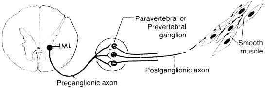
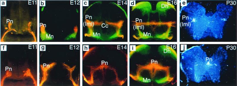
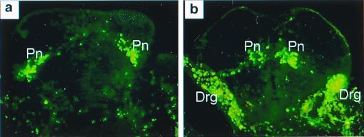
Mutation of the reeler gene (Reln) disrupts neuronal migration in several brain regions and gives rise to functional deficits such as ataxic gait and trembling in the reeler mutant mouse. Thus, the Reln product, reelin, is thought to control cell-cell interactions critical for cell positioning in the brain. Although an abundance of reelin transcript is found in the embryonic spinal cord [Ikeda, Y. & Terashima, T. (1997) Dev. Dyn. 210, 157-172; Schiffmann, S. N., Bernier, B. & Goffinet, A. M. (1997) Eur. J. Neurosci. 9, 1055-1071], it is generally thought that neuronal migration in the spinal cord is not affected by reelin. Here, however, we show that migration of sympathetic preganglionic neurons in the spinal cord is affected by reelin. This study thus indicates that reelin affects neuronal migration outside of the brain. Moreover, the relationship between reelin and migrating preganglionic neurons suggests that reelin acts as a barrier to neuronal migration.
Figures

Similar articles
-
Migration of sympathetic preganglionic neurons in the spinal cord of a C3G-deficient mouse suggests that C3G acts in the reelin signaling pathway.J Comp Neurol. 2012 Oct 1;520(14):3194-202. doi: 10.1002/cne.23086. J Comp Neurol. 2012. PMID: 22351125
-
Ectopic expression of reelin alters migration of sympathetic preganglionic neurons in the spinal cord.J Comp Neurol. 2009 Jul 10;515(2):260-8. doi: 10.1002/cne.22044. J Comp Neurol. 2009. PMID: 19412957
-
Evidence for a cell-specific action of Reelin in the spinal cord.Dev Biol. 2002 Apr 1;244(1):180-98. doi: 10.1006/dbio.2002.0580. Dev Biol. 2002. PMID: 11900467
-
Puzzling out the reeler brainteaser: does reelin signal to unique neural lineages?Brain Res. 2007 Apr 6;1140:41-50. doi: 10.1016/j.brainres.2006.02.056. Epub 2006 Mar 29. Brain Res. 2007. PMID: 16566902 Review.
-
[Developmental disorders and their responsible genes; the genes involved in neuronal positioning].No To Hattatsu. 2000 May;32(3):208-19. No To Hattatsu. 2000. PMID: 10824569 Review. Japanese.
Cited by
-
Reelin Functions, Mechanisms of Action and Signaling Pathways During Brain Development and Maturation.Biomolecules. 2020 Jun 26;10(6):964. doi: 10.3390/biom10060964. Biomolecules. 2020. PMID: 32604886 Free PMC article. Review.
-
Hormonal and morphological study of the pituitaries in reeler mice.Int J Exp Pathol. 2007 Jun;88(3):165-73. doi: 10.1111/j.1365-2613.2007.00528.x. Int J Exp Pathol. 2007. PMID: 17504446 Free PMC article.
-
Clinical and Neurobiological Relevance of Current Animal Models of Autism Spectrum Disorders.Biomol Ther (Seoul). 2016 May 1;24(3):207-43. doi: 10.4062/biomolther.2016.061. Biomol Ther (Seoul). 2016. PMID: 27133257 Free PMC article. Review.
-
Neuronal migration and the role of reelin during early development of the cerebral cortex.Mol Neurobiol. 2004 Dec;30(3):225-51. doi: 10.1385/MN:30:3:225. Mol Neurobiol. 2004. PMID: 15655250 Review.
-
Genetic and Epigenetic Etiology Underlying Autism Spectrum Disorder.J Clin Med. 2020 Mar 31;9(4):966. doi: 10.3390/jcm9040966. J Clin Med. 2020. PMID: 32244359 Free PMC article. Review.
References
Publication types
MeSH terms
Substances
LinkOut - more resources
Full Text Sources
Molecular Biology Databases
