. 2000 Aug 7;423(4):603-18.
doi: 10.1002/1096-9861(20000807)423:4<603::aid-cne6>3.0.co;2-f.

Distribution and development of nicotinic acetylcholine receptor subtypes in the optic tectum of Rana pipiens

Affiliations

Distribution and development of nicotinic acetylcholine receptor subtypes in the optic tectum of Rana pipiens

C M Butt et al. J Comp Neurol. .

Abstract

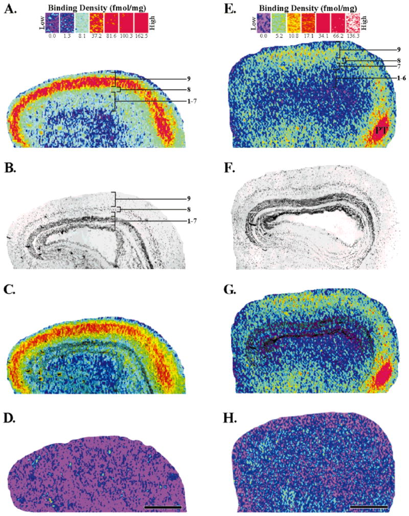
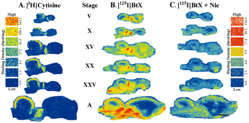
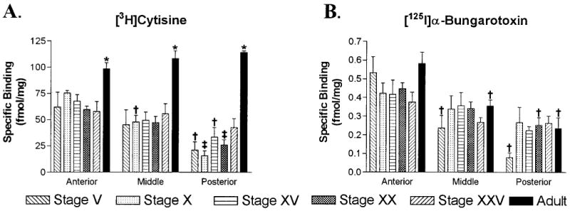
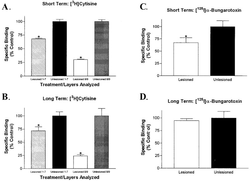
Acetylcholine allows the elicitation of visually evoked behaviors mediated by the frog optic tectum, but the mechanisms behind its effects are unknown. Although nicotinic acetylcholine receptors (nAChRs) exist in the tectum, their subtype has not been assessed. By using quantitative autoradiography, we examined the binding of [(3)H]cytisine and [(125)I]alpha-bungarotoxin in the laminated tectum. In mammalian systems, these radioligands bind with high affinity to alpha4 nAChR subunits and alpha7 nAChR subunits, respectively. [(3)H]Cytisine demonstrated high specific binding in adult frogs in retinorecipient layer 9, intermediate densities in layer 8, and low binding in layers 1-7 of the tectum. [(3)H]Cytisine binding was significantly higher in the tecta of adults than in those of tadpoles. Lesioning the optic nerve for 6 weeks decreased [(3)H]cytisine binding in layers 8/9 by 70+/-1%, whereas 6-month lesions decreased binding by 76+/-3%. Specific binding of [(125)I]alpha-bungarotoxin in adults was present only at intermediate levels in tectal layers 8 and 9, and undetectable in the deeper tectal layers. However, the nucleus isthmi, a midbrain structure reciprocally connected to the tectum, exhibited high levels of binding. There were no significant differences in tectal [(125)I]alpha-bungarotoxin binding between tadpoles and adults. Six-week lesions of the optic nerve decreased tectal [(125)I]alpha-bungarotoxin binding by 33+/-10%, but 6-month lesions had no effect. The pharmacokinetic characteristics of [(3)H]cytisine and [(125)I]alpha-bungarotoxin binding in the frog brain were similar to those demonstrated in several mammalian species. These results indicate that [(3)H]cytisine and [(125)I]alpha-bungarotoxin identify distinct nAChR subtypes in the tectum that likely contain non-alpha7 and alpha7 subunits, respectively. The majority of non-alpha7 receptors are likely associated with retinal ganglion cell terminals, whereas alpha7-containing receptors appear to have a different localization.


Figures

Fig. 1
Fig. 1
Schematic of a sagittal section through the adult optic tectum illustrating the location of the anterior, middle, and posterior tectal areas sampled in the developmental studies. Densitometry readings were taken from a 625-μm2 region in retinorecipient layer 9 for each of these areas. Scale bar = 500 μm.
Fig. 2
Fig. 2
Localization of [3H]cytisine (A,C) and [125I]α-bungarotoxin (E,G) binding sites in the adult optic tectum. The binding scales above each column provide a respective index of binding density (fmol/mg wet tissue weight) for each radioligand (see Materials and Methods for details). A: Specific [3H]cytisine (5 nM) binding in the optic tectum. The binding sites were most dense (red) in layer 9. Layer 8 had intermediate densities (yellow/green), whereas layers 1–7 had low densities (light green/light blue). Tectal layers were assigned by using the superimposed image seen in C. B: Brightfield image of the same tissue section that produced the autoradiogram (A). The optic tectum is a laminated structure consisting of alternating cellular and plexiform layers. The darkly stained, main cellular layer is layer 6. C: Superimposition of images A and B which accurately localizes the binding to specific, tectal layers. D: Nonspecific binding, as determined by the addition of nicotine (10 μM) to the incubation solution, was not detected when using [3H]cytisine at 5 nM. E: Total [125I]α-bungarotoxin binding (2.5 nM) in the optic tectum. The densest binding was seen in a pretectal area (PT). Intermediate binding densities were present in tectal layers 8 and 9. F: Brightfield image of the section that produced the autoradiogram in E. G: Superimposition of E and F localizes the binding to the tectal layers. H: Nonspecific [125I]α-bungarotoxin binding as determined by the addition of excess nicotine (10 μM). Anterior is to the right. Scale bars = 500 μm.
Fig. 3
Fig. 3
Schematic representation of a sagittal section through the frog brain that illustrates the general location and anatomical relationships of the brain areas depicted in Figure 4. Anterior is to the right.
Fig. 4
Fig. 4
Development and distribution of [3H]cytisine and [125I]α-bungarotoxin binding sites in early (stages V, X), middle (stage XV), and late (stages XX, XXV) tadpole and adult (A) frog optic tectum. A: [3H]Cytisine binding was primarily constrained to the superficial, tectal layers in tadpoles and in adults. Binding was present in every stage examined, although at early stages it was restricted to the rostral half of the tectum. Invasion of binding of the caudal half of the tectum was gradual and progressive with high binding densities achieved only in adulthood. Binding was also evident in the thalamus at all stages examined. Nonspecific binding of [3H]cytisine (5 nM) was not detected (not shown). B: [125I]α-Bungarotoxin (BtX) binding sites in the frog brain were distinct from those seen with [3H]cytisine. Consistent [125I]α-bungarotoxin binding was seen in the superficial tectal layers throughout development. This binding also had a pattern of expression that paralleled the tectal maturation gradient by first presenting itself in the rostral tectum (stages V, X) and then, later in development, in the caudal tectum as well (stages XV and above). The levels of both total and nonspecific binding in the tectum noticeably increased between stage XXV and adult animals. Binding was also observed in the telencephalon and throughout the thalamus and midbrain tegmentum at all stages of development. C: Nonspecific binding, as determined by competition with nicotine (Nic; 10 μM), represented 10% to 30% of the total [125I]α-bungarotoxin binding depending on the brain area sampled. Scale bar = 1 mm.
Fig. 5
Fig. 5
Quantitative analysis of [3H]cytisine and [125I]α-bungarotoxin binding in the retinorecipient layers of the tectum during development. A: Histograms displaying [3H]cytisine (5 nM) binding site densities for anterior, middle, and posterior regions of the optic tectum in tadpoles and adult frogs. Binding densities in every region of the adult tectum were equivalent and significantly higher than those found in any tadpole stage (*P < 0.02). In tadpoles, binding was present at fairly constant levels in the anterior tectum, at slightly lower levels in the middle tectum, and at still lower levels in the posterior tectum. Binding in the posterior tectum tended to increase with development. By stage XXV, binding densities in the posterior tectum were not significantly different from those in the anterior tectum. B: Histograms of [125I]α-bungarotoxin (2.5 nM) binding densities during development. Within the same age group, binding densities were greatest in the anterior tectum, intermediate in middle regions, and lowest in posterior ones. Within each of these regions, binding densities were equivalent across the different age groups. Error bars represent the standard error of the mean (S.E.M.; n = 4). † P < 0.05 compared to the anterior tectum of the same age, and ‡ P < 0.05 compared to both the anterior and middle tectum of the same age.
Fig. 6
Fig. 6
[125I]α-Bungarotoxin binding (2.5 nM) in the nucleus isthmi at different stages of development. Binding was clearly observable in the nucleus isthmi (arrows) of tadpole and adult tissue. In stages V, XX, XXV, and adults, binding densities were inhomogeneously distributed across this structure. A high level of nonspecific binding in stage X and stage XV tissue made it difficult to unambiguously identify binding sites with the nucleus isthmi (but see text). Comparisons of binding levels across the stages were not made because the results were from several individual experiments, and the treated tissue was not exposed to the same film. T, total [125I]α-bungarotoxin binding; B, brightfield image; O, overlay image for localization with the solid line outlining the nucleus isthmi; N, nonspecific binding with the dashed line outlining the nucleus isthmi. Anterior is to the right. Scale bar = 500 μm.
Fig. 7
Fig. 7
Effects of short and long term lesions of the optic nerve on [3H]cytisine (5 nM) and [125I]α-bungarotoxin (2.5 nM) binding site expression in the optic tectum. A: Representative autoradiogram of specific [3H]cytisine binding in a frontal section of the optic tectum after lesioning of the right optic nerve followed by a short (6-week) survival period. Binding in the deafferented (left) tectal lobe was dramatically reduced. B: [3H]Cytisine binding in the two tectal lobes after long term optic nerve lesion (6-month survival period). Residual binding in the superficial layers of the deafferented lobe was reduced even further by the extended survival time. C: Total [125I]α-bungarotoxin binding after lesioning and a 6-week survival time. Binding was marginally reduced in the deafferented (left) tectal lobe. D: Total [125I]α-bungarotoxin binding after a long-term lesion. No difference in binding was evident between the lesioned and unlesioned lobes. The four images should not be directly compared to one another because the experiments were performed at different times and exposed to different pieces of autoradiographic film. Quantitative analysis of these results is depicted in Figure 8. Scale bar = 1 mm.
Fig. 8
Fig. 8
Analysis of [3H]cytisine (5 nM) and [125I]α-bungarotoxin (2.5 nM) binding in the retinorecipient layers of the tectum after lesions of the adult optic nerve. A: Quantitative analysis of [3H]cytisine binding after lesioning of the adult optic nerve with a short survival period. The binding in layers 1–7 of the deafferented lobe was reduced by 31.5 ± 1.5%, whereas the binding in layers 8 and 9 was decreased by 70 ± 1%. B: Long survival periods after optic nerve lesions had a similar effect on [3H]cytisine binding in the deafferented lobe. Binding in layers 1–7 was decreased by 28 ± 8% and that in layers 8 and 9 was reduced by 76 ± 3%. C: Short-term optic nerve lesions had a small, but significant, effect on specific [125I]α-bungarotoxin binding in the superficial layers of the deafferented lobe. Binding was reduced by 33 ± 10% in layers 8 and 9. D: Long-term optic nerve lesions did not affect specific [125I]α-bungarotoxin binding in tectal layers 8 and 9. There was no significant difference between afferented and deafferented lobes. Error bars represent the S.E.M. (*P < 0.05, paired, two-tailed t-test; n = 4 or 5).
Fig. 9
Fig. 9
Pharmacological analysis of [3H]cytisine and [125I]α-bungarotoxin binding in layers 8 and 9 of the adult optic tectum. A: Saturation binding analysis of [3H]cytisine binding. The apparent dissociation constant (KD) was 0.47 ± 0.04 nM with a maximum binding capacity (Bmax) of 40.4 ± 0.71 fmol/mg. The data were best fit by an equation for binding to a single site. The inset is a Scatchard plot of the same data that yielded similar binding constants. B: Competition binding analysis of [3H]cytisine (5 nM) binding. Dihydro-β-erythroidine (DHBE), which is a competitive antagonist at non-α7 nicotinic acetylcholine receptors (nAChRs) in mammalian systems, competed with [3H]cytisine at two sites. The high affinity DHBE site demonstrated an inhibition constant (Ki) of 40.3 pM, whereas the low-affinity site had a Ki of 24.5 nM. Methyllycaconitine (MLA) and unlabeled α-bungarotoxin (BtX) are both high-affinity, competitive antagonists of the α7 nAChR in mammalian systems and did not compete with [3H]cytisine binding until micromolar concentrations of competitor were present. MLA had a Ki of 3.29 μM. Unlabeled BtX was unable to reduce [3H]cytisine binding to background levels and demonstrated a Ki of 12.5 μM. C: Saturation analysis of [125I]α-bungarotoxin binding demonstrated a KD = 0.81 ± 0.19 nM and a Bmax = 1.86 ± 0.10 fmol/mg. The data fit best to an equation for single-site binding, and Scatchard analysis (inset) again produced similar binding values. D: Competition of MLA, unlabeled cytisine, and DHBE against [125I]α-bungarotoxin (2.5 nM) binding. The competition curves fit to equations that produced the following inhibition constants: MLA = 2.3 pM and 2.6 nM, unlabeled cytisine = 17.6 nM, DHBE = 21.1 μ M. All points are the mean ± S.E.M. of specific binding (n = 4 or 5) for all graphs.

Similar articles

Cited by

References

    1. Alkondon M, Pereira EF, Eisenberg HM, Albuquerque EX. Choline and selective antagonists identify two subtypes of nicotinic acetylcholine receptors that modulate GABA release from CA1 interneurons in rat hippocampal slices. J Neurosci. 1999;19:2693–2705. - PMC - PubMed
    1. Anand R, Peng X, Ballesta JJ, Lindstrom J. Pharmacological characterization of alpha-bungarotoxin-sensitive acetylcholine receptors immunoisolated from chick retina: contrasting properties of alpha 7 and alpha 8 subunit-containing subtypes. Mol Pharmacol. 1993;44:1046–1050. - PubMed
    1. Aubert I, Cecyre D, Gauthier S, Quirion R. Comparative ontogenic profile of cholinergic markers, including nicotinic and muscarinic receptors, in the rat brain. J Comp Neurol. 1996;369:31–55. - PubMed
    1. Barrantes GE, Rogers AT, Lindstrom J, Wonnacott S. alpha-bungarotoxin binding sites in rat hippocampal and cortical cultures: initial characterisation, colocalisation with alpha 7 subunits and up-regulation by chronic nicotine treatment. Brain Res. 1995;672:228–236. - PubMed
    1. Bear MF, Singer W. Modulation of visual cortical plasticity by acetylcholine and noradrenaline. Nature. 1986;320:172–176. - PubMed

Publication types

LinkOut - more resources