

doi: 10.1038/75869.
The structure of the transcriptional antiterminator NusB from Escherichia coli
Affiliations
- PMID: 10881193
- PMCID: PMC8397614
- DOI: 10.1038/75869
Item in Clipboard
The structure of the transcriptional antiterminator NusB from Escherichia coli
Nat Struct Biol.
2000 Jun.
Abstract
We have determined the solution structure of NusB, a transcription antitermination protein from Escherichia coli. The structure reveals a novel, all alpha-helical protein fold. NusB mutations that cause a loss of function (NusB5) or alter specificity for RNA targets (NusB101) are localized to surface residues and likely affect RNA-protein or protein-protein interactions. Residues that are highly conserved among homologs stabilize the protein core. The solution structure of E. coli NusB presented here resembles that of Mycobacterium tuberculosis NusB determined by X-ray diffraction, but differs substantially from a solution structure of E. coli NusB reported earlier.
Figures
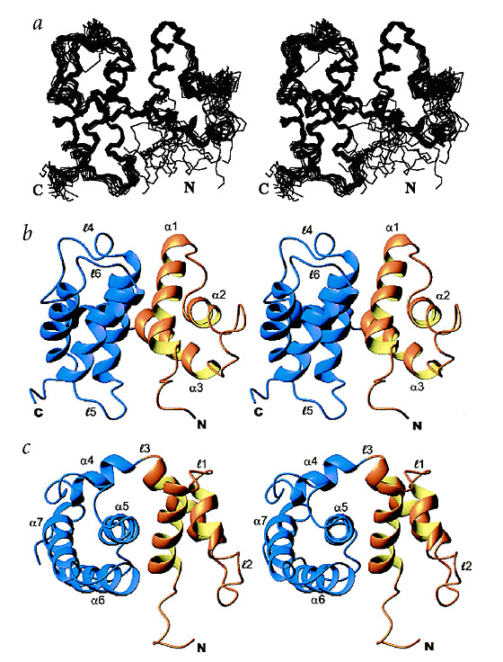
a, Stereo view showing the superposition of 15 of the lowest energy structures of NusB. b, Stereo view of a ribbon trace of NusB. The N-terminal subdomain is colored gold, and the C-terminal subdomain is colored purple. Helices and loops are labeled as discussed in the text. c, Stereo view showing a 90y° rotation of (b).
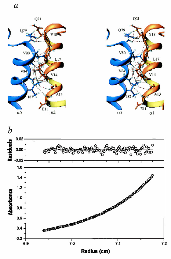
a, Long range NOE contacts between α1 (gold) and α5 (blue). These NOEs define the orientation of the N-terminal and C-terminal subdomains relative to each other. b, The determination of the molecular mass of E. coli NusB in 50 mM potassium phosphate, pH 6.8, 50 mM NaCl and 1 mM DTT at 20 °C. The absorbance gradient (at 280 nm) in the centrifuge cell after attaining sedimentation equilibrium at 25,000 r.p.m. is shown in the bottom panel. The solid line is the result of fitting to a single ideal species and the open circles are the experimental values. The corresponding top panel shows the difference between the fit and the experimental values as a function of radial position (residuals).

a, Sequence alignment of NusB orthologs. Amino acids identical to the E. coli sequence are highlighted in green. Amino acids that represent a conservative substitution from the E. coli sequence are highlighted in orange. Amino acids that are identical across all NusB sequences are marked by a solid arrow. Amino acid positions with only conserved substitutions across all sequences are marked by an open arrow. b, Localization of conserved residues on NusB. The side chains of conserved or identical residues across all NusB proteins are displayed and labeled.
References
-
- Greenblatt J. Transcriptional regulation. 1992. pp. 203–226.
MeSH terms
Substances
Associated data
- Actions
LinkOut - more resources
Full Text Sources
Molecular Biology Databases