. 2000 Jul 15;20(14):5292-9.
doi: 10.1523/JNEUROSCI.20-14-05292.2000.

Modulation of interleukin-1beta and tumor necrosis factor alpha signaling by P2 purinergic receptors in human fetal astrocytes

Affiliations

Modulation of interleukin-1beta and tumor necrosis factor alpha signaling by P2 purinergic receptors in human fetal astrocytes

J S Liu et al. J Neurosci. .

Abstract

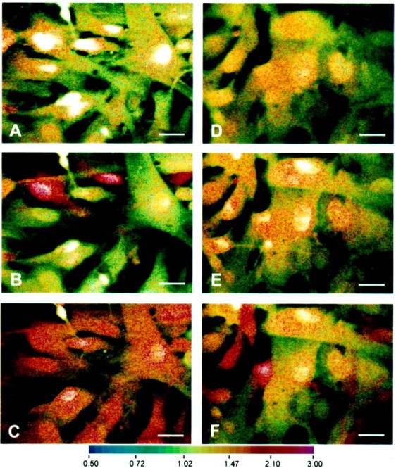
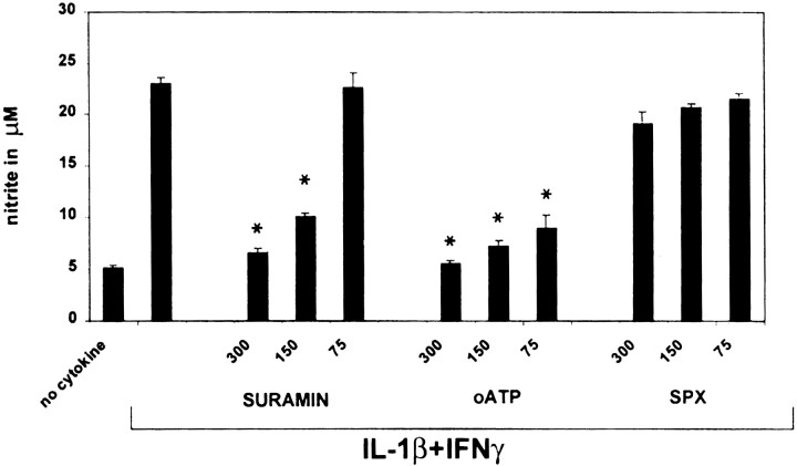
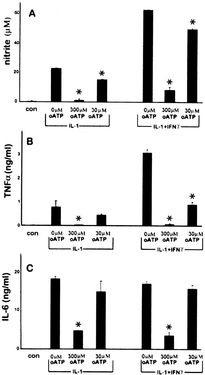
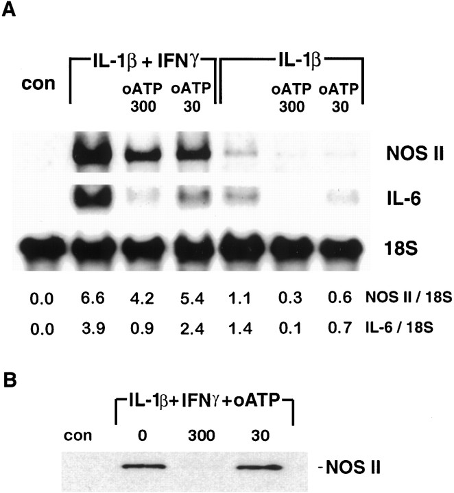
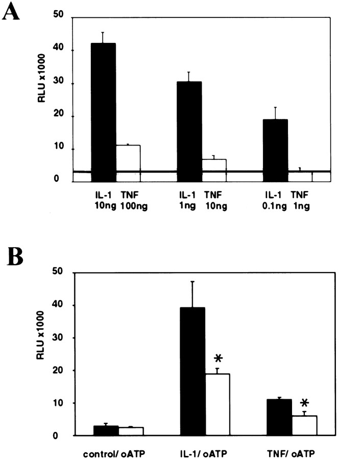
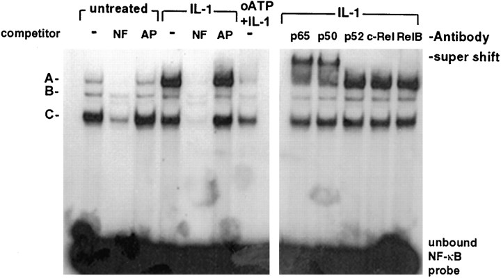
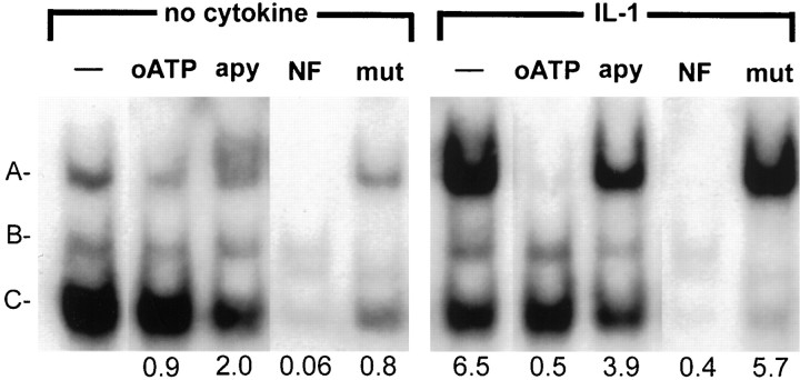
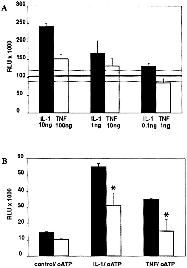
In human astrocytes, interleukin-1beta (IL-1beta) is a potent inducer of genes associated with inflammation. In this study, we tested the hypothesis that in primary cultures of human fetal astrocytes signaling by the P2 purinergic nucleotide receptor pathway contributes to, or modulates, cytokine-mediated signal transduction. Calcium imaging studies indicated that most cells in culture responded to ATP, whereas only a subpopulation responded to UTP. Pretreatment of astrocytes with P2 receptor antagonists, including suramin and periodate oxidized ATP (oATP), resulted in a significant downregulation of IL-1beta-stimulated expression of nitric oxide, tumor necrosis factor (TNFalpha), and IL-6 at both the protein and mRNA levels, without affecting cell viability. In cells transiently transfected with reporter constructs, IL-1beta demonstrated more potent activation of the transcription factors nuclear factor -kappaB (NF-kappaB) and activator protein-1 (AP-1) than TNFalpha. However, pretreatment with oATP downregulated activation of NF-kappaB and AP-1 by IL-1beta or TNFalpha. Electromobility shift assays using oligonucleotides containing specific NF-kappaB binding sequences confirmed that pretreatment with oATP or apyrase attenuated cytokine-mediated induction of this transcription factor. From these data, we conclude that P2 receptor-mediated signaling intersects with that of IL-1beta and TNFalpha to regulate responses to cytokines in the CNS. Because inflammation, trauma, and stress all lead to the release of high levels of extracellular nucleotides, such as ATP and UTP, signaling via P2 receptors may provide a mechanism whereby cells can sense and respond to events occurring in the extracellular environment and can fine tune the transcription of genes involved in the inflammatory response.


Figures

Fig. 1.
Fig. 1.
Exogenously added nucleotides results in transient elevations of intracellular calcium in human astrocytes that is blocked by oATP. Astrocytes were loaded with 10 μm Indo-1 for 45 min and visualized by real-time confocal imaging microscopy. Increase in intracellular calcium was visualized as a shift toward the red spectrum. A–C show the same field without stimulation (A) and after exposure to 100 μmUTP (B) or 100 μm ATP (C). D–F are also of the same field in cultures pretreated with 300 μm oATP, showing control no stimulation (D) and 100 μm UTP (E) or 100 μmATP (F). Note the similarity in basal intracellular calcium levels in control (A) and oATP-treated (D) cells. All panelsof nucleotide-treated cultures are shown at mean peak intracellular calcium concentrations after treatments. The experiment shown is one of three, each on cells derived from different brains. Scale bars, 25 μm.
Fig. 2.
Fig. 2.
Blockade of P2 receptors on astrocytes downregulates nitrite production. Astrocytes were pretreated for 2 hr with the inhibitors oATP, suramin, and SPX, at concentrations of 300, 150, and 75 μm. oATP was washed out before the addition of IL-1β (10 ng/ml) plus IFNγ (200 U/ml). Nitrite was determined at 48 hr. The P2 inhibitors oATP and suramin demonstrated a dose-dependent inhibition of nitrite production. In contrast, the P1 inhibitor SPX had no effect. *p < 0.05 indicates statistically significant reduction of nitrite compared with cytokine stimulation without inhibitors. Data represent the mean and SD of astrocytes from five different wells per data point and are representative of four separate experiments on cells derived from different brains.
Fig. 3.
Fig. 3.
Blockade of P2 receptors on astrocytes downregulates induction of TNFα, IL-6, and nitrite production. Astrocytes were pretreated with varying doses of oATP (0, 300, or 30 μm) for 2 hr, washed, and exposed to IL-1β(10 ng/ml) or IL-1β plus IFNγ (200 U/ml) as indicated. Control cultures (con) consisted of astrocytes treated with oATP alone (300 μm) without cytokines. TNFα (B) and IL-6 (C) levels in the supernatant were determined at 16 hr and nitrite (A) at 48 hr. Pretreatment with oATP resulted in a dose-dependent inhibition of nitrite, TNFα, and IL-6 after stimulation with IL-1β alone or in combination with IFNγ. Note also that, whereas IFNγ potently synergized with IL-1β for nitrite and TNFα production, it had little effect on IL-6 expression. *p < 0.05 indicates statistically significant reduction compared with cytokine stimulation without inhibitors. Data represent the mean and SD of results from three different wells and are representative of cells derived from three different brains.
Fig. 4.
Fig. 4.
Blockade of astrocyte P2 receptors results in inhibition of NOS II, and IL-6 mRNA and NOS II protein expression.A, RNA was extracted 24 hr after cytokine treatment from the same cultures described in Figure 3 and subjected to Northern analysis of NOS II, IL-6, and 18 S mRNA (A). Densitometric ratios of NOS II and IL-6 to 18 S are givenbelow each lane. The results show that P2 blockade acts at the level of mRNA expression. Data shown are representative of three separate experiments on cells derived from three different brains. B, NOS II protein expression was determined by Western blot analysis of homogenates. Astrocytes were pretreated with 0, 30, or 300 μm oATP for 2 hr, washed, and treated with IL-1β (10 ng/ml) plus IFNγ (200 U/ml) for 48 hr. Cytokine stimulation resulted in strong induction of NOS II (130 kDa band) that was markedly reduced in cells that had been pretreated with 300 μm oATP. Data shown are representative of four independent experiments.
Fig. 5.
Fig. 5.
IL-1β and TNFα activation of NF-κB is dose-dependent and downregulated by P2 receptor blockade.A, Astrocyte cultures were transiently transfected with a reporter construct for NF-κB activity. Cells were treated with either IL-1β (0.1, 1, or 10 ng/ml) or TNFα (1, 10, or 100 ng/ml), and luciferase activity was measured at 5 hr (A). Level of luciferase activity (expressed as RLU) in untreated cells is represented by the transverse line, and SDs of these background levels are demarcated by the dotted lines. NF-κB induction by both IL-1β and TNFα was dose-dependent. However, IL-1β treatment resulted in much higher levels of NF-κB activation. Data shown represent the mean and SD of three separate measurements and are representative of three separate experiments derived from cells from three different brains. B, Cells were transfected as above and treated with both IL-1 (10 ng/ml) and TNFα (100 ng/ml) in the presence or absence of pretreatment with oATP (300 μm). As before, IL-1 and TNFα resulted in induction of NF-κB (with IL-1 induction greater than TNFα induction), and this was downregulated by pretreatment using oATP blockade of P2 receptors, which had no effect on background levels of NF-κB activation. Data shown are the mean and SD of three separate measurements and are representative of three separate experiments with cells from three different brains.
Fig. 6.
Fig. 6.
IL-1β induction of NF-κB binding is downregulated by blockade of P2 receptor signaling. Astrocyte cultures were treated with IL-1β in the presence or absence of pretreatment with oATP. Nuclei were harvested at 30 min and subjected to EMSA with a radiolabeled oligonucleotide probe containing the NF-κB binding site along with specific (lane NF) and nonspecific (lane AP) competitor oligonucleotides. Theleft shows control samples, IL-1β-treated samples, and IL-1β-treated samples after oATP pretreatment. Three shift complexes were observed (A–C) in control cells. Complex A was strongly induced by IL-1β and downregulated by oATP pretreatment. The right panel shows the results of EMSA on IL-1β-treated astrocytes incubated with specific antibodies for NF-κB family members. Only incubation with antibodies to p50 and p65 resulted in the formation of a supershift complex. The experiment shown is representative of four separate experiments using astrocytes derived from four different brains.
Fig. 7.
Fig. 7.
IL-1β induction of NF-κB binding is downregulated by breakdown of extracellular nucleotides by apyrase. Astrocyte cultures were treated with IL-1β in the presence or absence of apyrase (lane apy; 20 U/ml) pretreatment for 30 min or oATP (lane oATP) pretreatment as described previously. Nuclei were harvested at 30 min and subjected to EMSA with a radiolabeled oligonucleotide probe containing the NF-κB binding site along with wild-type (lane NF) and mutant (lane mut) competitor oligonucleotides. Control samples, IL-1β-treated samples, and IL-1β-treated samples after apyrase pretreatment or oATP are shown. Three shift complexes were observed (A–C) in control cells. Complex A was strongly induced by IL-1β and downregulated by oATP pretreatment, as well as by apyrase. Quantitative analysis of these data is shownbelow each lane and represents the densitometric ratios for complex A compared with the value for the complex A in the control sample (left lane). Data shown are representative of two separate experiments with cells from two different brains.
Fig. 8.
Fig. 8.
IL-1β and TNFα activation of AP-1 is dose-dependent and downregulated by P2 receptor blockade.A, Astrocyte cultures were transiently transfected with a reporter construct for AP-1 activity. Cells were treated with either IL-1β (0.1, 1, or 10 ng/ml) or TNFα (1, 10, or 100 ng/ml), and luciferase activity was measured in RLU after 4–5 hr (A). Levels of luciferase activity (expressed as RLU) in untreated cells is represented by the transverse line, and SDs of these background levels are demarcated by thedotted lines. AP-1 induction by both IL-1β and TNFα was dose-dependent. However, IL-1β treatment resulted in higher levels of AP-1 activation. B, cells were transfected as above and treated with either IL-1β (10 ng/ml) or TNFα (100 ng/ml) in the presence or absence of pretreatment with oATP as described above. As before, IL-1 and TNFα resulted in induction of AP-1 (with IL-1β greater than TNFα) that was downregulated by pretreatment with oATP. Blockade of P2 receptors with oATP had no effect on background levels of AP-1 activation. Data shown are representative of three separate experiments on cells from three different brains.

Similar articles

Cited by

References

    1. Akama KT, Albanese C, Pestell RG, Van Eldik LJ. Amyloid beta-peptide stimulates nitric oxide production in astrocytes through an NFkappaB-dependent mechanism. Proc Natl Acad Sci USA. 1998;95:5795–5800. - PMC - PubMed
    1. Brambilla R, Burnstock G, Bonazzi A, Ceruti S, Cattabeni F, Abbracchio MP. Cyclo-oxygenase-2 mediates P2Y receptor-induced reactive astrogliosis. Br J Pharmacol. 1999;126:563–567. - PMC - PubMed
    1. Breder CD, Dinarello CA, Saper CB. Interleukin-1 immunoreactive innervation of the human hypothalamus. Science. 1988;240:321–324. - PubMed
    1. Bruner G, Murphy S. Purinergic P2Y receptors on astrocytes are directly coupled to phospholipase A2. Glia. 1993;7:219–224. - PubMed
    1. Burnstock G. The past, present and future of purine nucleotides as signalling molecules. Neuropharmacology. 1997;36:1127–1139. - PubMed

Publication types

MeSH terms