Comparative Study
. 2000 Jul 5;97(14):7732-7.
doi: 10.1073/pnas.97.14.7732.

Crystal structure of human stem cell factor: implication for stem cell factor receptor dimerization and activation

Affiliations
Comparative Study

Crystal structure of human stem cell factor: implication for stem cell factor receptor dimerization and activation

Z Zhang et al. Proc Natl Acad Sci U S A. .

Abstract

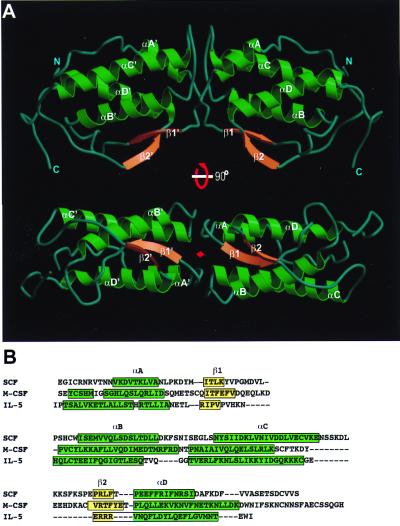
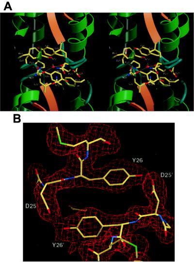
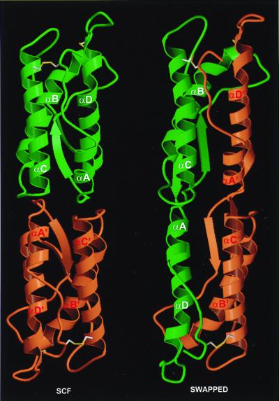
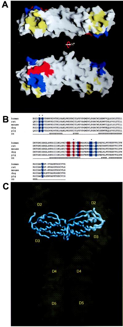
Stem cell factor (SCF) plays important roles in hematopoiesis and the survival, proliferation, and differentiation of mast cells, melanocytes, and germ cells. SCF mediates its biological effects by binding to and activating a receptor tyrosine kinase designated c-kit or SCF receptor. In this report we describe the 2.3-A crystal structure of the functional core of recombinant human SCF. SCF is a noncovalent homodimer composed of two slightly wedged protomers. Each SCF protomer exhibits an antiparallel four-helix bundle fold. Dimerization is mediated by extensive polar and nonpolar interactions between the two protomers with a large buried surface area. Finally, we have identified a hydrophobic crevice and a charged region at the tail of each protomer that functions as a potential receptor-binding site. On the basis of these observations, a model for SCF small middle dotc-kit complex formation and dimerization is proposed.


Figures

Figure 1
Figure 1
Overall structure of SCF and its relationship with other cytokines. (A) Ribbon representation of the SCF structure, in two views related by a rotation of approximately 90°. The termini and secondary structures are labeled; the β-strands are rendered as orange arrows, the helices as green ribbons, and the loop regions as gray tubes. The twofold axis is marked with a red diamond. (B) Sequence alignment based on secondary structures of SCF, M-CSF, and IL-5. Secondary structure assignments for M-CSF and IL-5 are from the Protein Data Bank. β-Strands are yellow and helices are bright green.
Figure 2
Figure 2
Dimer interface. (A) Stereoview of the dimer interface. For clarity, only side chains of residues at the core of the interface are shown. Color coding of the secondary structures is the same as in Fig. 1A. (B) 2FoFc electron density, contoured at 1.2σ, for the hydrogen bond circle of Tyr-26 and Asp-25′ at the dimer interface.
Figure 3
Figure 3
Model of covalent SCF dimer. The noncovalent (native) dimer is on the left and a model for the covalent SCF dimer is on the right. Each protomer is colored either orange or green. The disulfide bonds are shown in ball-and-stick with sulfur atoms colored yellow.
Figure 4
Figure 4
A potential binding site on SCF for c-kit and a model of SCF⋅SCFR complex. (A) Molecular surface of SCF and proposed c-kit binding regions, in two views related by a rotation of approximately 90°. A hydrophobic crevice at both tails is colored yellow. Two basic patches are colored blue, and the acidic patch is colored red. (B) Sequence alignments of human, rat, mouse, dog, and pig SCFs. Residues of the acidic patch are colored red and residues of the two basic patches are colored blue. Asterisks mark amino acid residues that are altered in rodents. The secondary structures (SS) are marked below the sequences with H representing helices and E representing β-strands. (C) Proposed model of the SCF in complex with Ig-like domains 2–5 of the extracellular domain of c-kit (labeled D2 to D5). SCF dimer is represented in a worm model, and the c-kit model is represented by a molecular surface.

References

    1. Galli S J, Zsebo K M, Geissler E N. Adv Immunol. 1994;55:1–96. - PubMed
    1. Williams D E, Eisenman J, Baird A, Rauch C, Van Ness K, March C J, Park L S, Martin U, Mochizuki D Y, Boswell H S, et al. Cell. 1990;63:167–174. - PubMed
    1. Zsebo K M, Williams D A, Geissler E N, Broudy V C, Martin F H, Atkins H L, Hsu R Y, Birkett N C, Okino K H, Murdock D C, et al. Cell. 1990;63:213–224. - PubMed
    1. Huang E, Nocka K, Beier D R, Chu T Y, Buck J, Lahm H W, Wellner D, Leder P, Besmer P. Cell. 1990;63:225–233. - PubMed
    1. Lemmon M A, Schlessinger J. Trends Biochem Sci. 1994;19:459–463. - PubMed

Publication types

Associated data

LinkOut - more resources