. 2000 Jul 5;97(14):7784-9.
doi: 10.1073/pnas.97.14.7784.

Escherichia coli CspA-family RNA chaperones are transcription antiterminators

Affiliations

Escherichia coli CspA-family RNA chaperones are transcription antiterminators

W Bae et al. Proc Natl Acad Sci U S A. .

Abstract

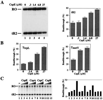
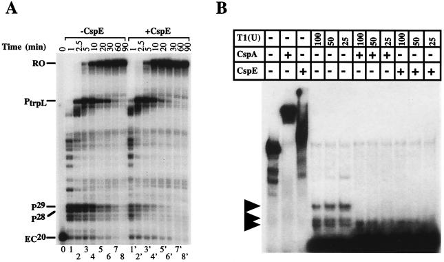
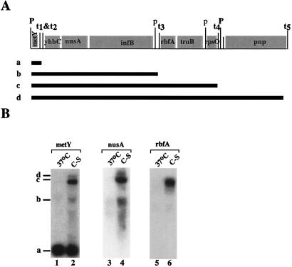
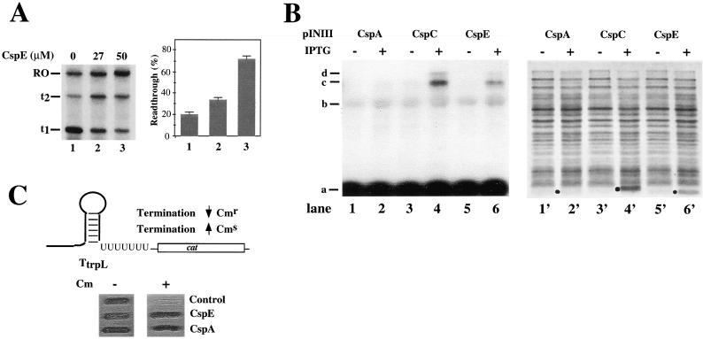
CspA, the major cold-shock protein of Escherichia coli, is an RNA chaperone, which is thought to facilitate translation at low temperature by destabilizing mRNA structures. Here we demonstrate that CspA, as well as homologous RNA chaperones CspE and CspC, are transcription antiterminators. In vitro, the addition of physiological concentrations of recombinant CspA, CspE, or CspC decreased transcription termination at several intrinsic terminators and also decreased transcription pausing. In vivo, overexpression of cloned CspC and CspE at 37 degrees C was sufficient to induce transcription of the metY-rpsO operon genes nusA, infB, rbfA, and pnp located downstream of multiple transcription terminators. Similar induction of downstream metY-rpsO operon genes was observed at cold shock, a condition to which the cell responds by massive overproduction of CspA. The products of nusA, infB, rbfA, and pnp-NusA, IF2, RbfA, and PNP-are known to be induced at cold shock. We propose that the cold-shock induction of nusA, infB, rbfA, and pnp occurs through transcription antitermination, which is mediated by CspA and other cold shock-induced Csp proteins.


Figures

Figure 1
Figure 1
Csp proteins act as transcription antiterminators in vitro. (A) CspE induces transcription antitermination on the tR2 terminator. Transcription elongation complexes artificially stalled at position 20 (EC20) were prepared by using a DNA template containing the T7 A1 promoter fused to tR2 terminator and αHisRNAP immobilized on Ni2+-nitrilotriacetic acid agarose. The indicated concentrations of recombinant CspE were added to washed EC20, and transcription elongation was resumed by the addition of NTP. Reaction products were separated by denaturing PAGE and visualized by autoradiography. RO and tR2 indicate the runoff and the tR2-terminated transcripts, respectively. The gel shown (Left) was quantified by using phosphorimagery, and the readthrough efficiency, RE, was calculated (RE = {[RO transcripts]/[total transcripts (tR2 + RO)]} × 100%). The results are schematically presented (Right). (B) CspE induces transcription antitermination at the trpL and metY terminators. The in vitro transcription and product analysis were carried out under the same condition as described in A, except the template used contained TtrpL (Left) or TmetYt1 (Right) fused to the T7 A1 promoter. (C) Effects of CspA, CspB, CspC, and CspE on transcription termination at tR2. Csp proteins (CspE, CspA, CspB, and CspC) were used for in vitro transcription assays, as described in A: lane 1, no Csp protein; 1.4 μM Csp for lanes 2, 5, 8, and 11; 10 μM Csp for lanes 3, 6, 9, and 12; and 20 μM Csp for lanes 4, 7, 10, and 13. The RE values were quantified and are schematically presented (Right).
Figure 2
Figure 2
Effect of CspE on transcription pausing, and RNA chaperoning activity of Csp proteins. (A) In vitro transcription experiment was performed by using DNA fragment consisting of the T7 A1 terminator fused to the trpL pause site (PtrpL) as template. To observe pausing, low concentration of NTP (5 μM) was used for chasing the primary EC (EC20). Reactions were terminated at the times indicated. (B) Stimulation of RNase activity by CspE and CspA. Ribonuclease assays were carried out as described in Materials and Methods. The RNA substrate used is the same as used previously for the ribonuclease experiment for CspA (6). RNase T1 was added as indicated (Top) in units. Lane 1, RNA substrate alone; lane 2, RNA substrate plus CspA (27 μM); lane 3, RNA substrate plus CspE (27 μM); lanes 4–6, 100, 50, 25 unit of RNase T1, respectively; lanes 7–9, the same as lanes 4–6, respectively, except that CspA (27 μM) was added before adding RNase T1; lanes 10–12, the same as lanes 4–6, respectively, except that CspE (27 μM) was added before adding RNase T1. The reaction mixtures were incubated on ice for 10 min and loaded onto the 8% acrylamide gel.
Figure 3
Figure 3
The downstream genes of the metY-rpsO operon are induced by cold shock. (A) The gene structure of the metY and rpsO operons (33), which contains metY (tRNAf2), yhbC (an unidentified ORF), nusA (NusA), infB(IF2), rbfA (RbfA, 34), truB (P35, E. coli Psi synthase), rpsO (S15), and pnp (PNPase). The positions of four intercistronic intrinsic terminators from t1 to t4 and promoters are indicated. Capital “P”s represent promoters that are relatively strong. Lowercase “p”s represent weak promoters. The hypothetical transcripts, which are initiated from the metY promoter but terminated at different terminators sequentially, are shown and indicated with letters, “a–d”. (B) RNA was isolated from cells grown at 37°C and cold-shocked cells (C-S). An equal amount of RNA (20 μg) was loaded in each lane. Northern blot analysis was carried out by using the metY gene as a probe (Left). After the membrane was washed, it was reprobed with the rbfA gene (Center) and the nusA gene (Right). The transcripts are indicated with the letters “a–d,” whose identities are shown in A.
Figure 4
Figure 4
Csp proteins cause transcription antitermination at the metY-rpsO operon in vitro and in vivo. (A) A DNA fragment containing two consecutive intrinsic terminators, t1 and t2, found downstream of metY and genetically fused to the T7 A1 promoter, was used as a template to examine the effect of CspE on the termination at these terminators. The experiment was performed and data analyzed as described in the Fig. 1 legend. The combined RE values at t1 and t2 were calculated (RE = {[RO transcripts]/[total transcripts (t1+ t2+ RO)]} × 100%) and are graphically presented (Right). (B) E. coli cells were transformed with the pINCspA, pINCspC, or pINCspE plasmids, grown at 37°C to OD600 of 0.3–0.4, and csp expression was induced by the addition of 1 mM IPTG. After 60 min incubation at 37°C, half of the culture was subjected to the isolation of total RNA and Northern blot analysis by using the metY gene as a probe (lanes 1–6), whereas the other half was analyzed by SDS/PAGE on a 17.5% gel (lanes 1′-6′). The overexpressed proteins are indicated by black dots. (C) RL211 E. coli cells containing a cat gene cassette positioned downstream of the trpL terminator (36) were transformed with pINCspA, pINCspC, or control pINIII, and spotted on plates containing 30 μg/ml chloramphenicol and1 mM IPTG. The results of overnight cell growth are presented.

References

    1. Yamanaka K, Fang L, Inouye M. Mol Microbiol. 1998;27:247–255. - PubMed
    1. Wolffe A P. Bioassays. 1994;16:245–251. - PubMed
    1. Graumann P, Wendrich T M, Weber M H W, Schroder K, Marahiel M A. Mol Microbiol. 1997;25:741–756. - PubMed
    1. Bae W, Jones P G, Inouye M. J Bacteriol. 1997;179:7081–7088. - PMC - PubMed
    1. Bae W, Phadtare S, Severinov K, Inouye M. Mol Microbiol. 1999;31:1429–1442. - PubMed

Publication types

MeSH terms

LinkOut - more resources