. 2000 Jul 5;97(14):7871-6.
doi: 10.1073/pnas.97.14.7871.

Selective depletion of heat shock protein 70 (Hsp70) activates a tumor-specific death program that is independent of caspases and bypasses Bcl-2

Affiliations

Selective depletion of heat shock protein 70 (Hsp70) activates a tumor-specific death program that is independent of caspases and bypasses Bcl-2

J Nylandsted et al. Proc Natl Acad Sci U S A. .

Abstract

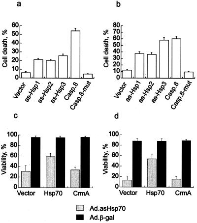
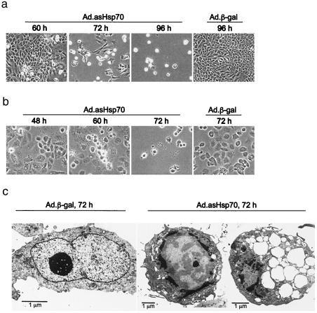
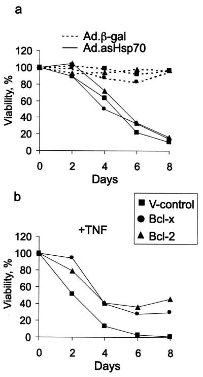
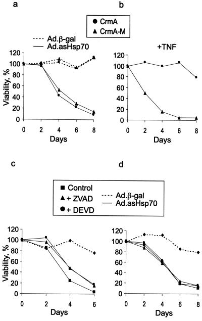
Heat shock protein 70 is an antiapoptotic chaperone protein highly expressed in human breast tumors and tumor cell lines. Here, we demonstrate that the mere inhibition of its synthesis by adenoviral transfer or classical transfection of antisense Hsp70 cDNA (asHsp70) results in massive death of human breast cancer cells (MDA-MB-468, MCF-7, BT-549, and SK-BR-3), whereas the survival of nontumorigenic breast epithelial cells (HBL-100) or fibroblasts (WI-38) is not affected. Despite the apoptotic morphology as judged by electron microscopy, the asHsp70-induced death was independent of known caspases and the p53 tumor suppressor protein. Furthermore, Bcl-2 and Bcl-X(L), which protect tumor cells from most forms of apoptosis, failed to rescue breast cancer cells from asHsp70-induced death. These results show that tumorigenic breast cancer cells depend on the constitutive high expression of Hsp70 to suppress a transformation-associated death program. Neutralization of Hsp70 may open new possibilities for treatment of cancers that have acquired resistance to therapies activating the classical apoptosis pathway.


Figures

Figure 1
Figure 1
Inhibition of Hsp70 synthesis results in decreased survival of breast cancer cells. The indicated human breast carcinoma cell lines (ad), lung-derived fibroblasts (e), and breast-derived nontumorigenic cells (f) were infected with Ad.asHsp70 or Ad.β-gal. The survival of cells was analyzed at indicated times after infection by counting the cells (ad and f) or the MTT assay (e). The survival of Ad.β-gal-infected cells is expressed as a percentage compared with noninfected cells and that of Ad.asHsp70-infected cells as a percentage compared with Ad.β-gal-infected cells. The columns represent means of cell counts from a minimum of eight fields (ad and f) or triplicate MTT assay measurements (e) ± SD. Proteins (20 μg per lane) from similarly infected cell lysates were analyzed for Hsp70, Hsc70, and glyceraldehyde-3-phosphate dehydrogenase by immunoblotting. All experiments were repeated one to three times with essentially the same results.
Figure 2
Figure 2
Antisense Hsp70 DNA induces cell death in breast cancer cells and Ad.asHsp70-infected cells can be rescued by overexpression of human Hsp70. Subconfluent MCF-7 (a) or MDA-MB-468 (b) cells were transfected with pcDNA-3 vector or the same vector with three different antisense Hsp70 sequences (as-Hsp1, -2, or -3) or the entire coding sequence for caspase 8 (Casp.8) or mutated caspase 8 (Casp.8-mut). The successfully transfected cells were visualized by cotransfection (1:10) of pEGFP-N1, and the percentage of green dead cells of all green cells was counted 96 h after transfection. Rescue experiments were performed in a similar manner by cotransfecting pEGFP-N1 with an empty vector, pSV-hsp70-tag or pcDNA3-CrmA into MCF-7 (c) or MDA-MB-468 (d) cells 24 h after the infection with indicated viruses. The percentage of green-surviving cells of all green cells was counted 96 h after the infection. The values represent means of five fields each with a minimum of 100 cells ± SD. The experiments were repeated twice with essentially the same results.
Figure 3
Figure 3
Breast cancer cells infected with Ad.asHsp70 display an apoptotic morphology. MCF-7 (a and c) and MDA-MB-468 (b) breast cancer cells were infected with indicated adenoviruses and analyzed for morphological changes by phase contrast microscopy (a and b) or electron microscopy (c) at indicated times after the infection.
Figure 4
Figure 4
Bcl-2 and Bcl-XL fail to protect breast cancer cells from Ad.asHsp70-induced cell death. Subconfluent MCF-7 cells transfected with an empty vector (V-control) or those overexpressing Bcl-2 or Bcl-XL were infected with indicated viruses (a) or treated with 10 ng/ml of TNF (b). The survival of the cells as compared with noninfected cells was analyzed by MTT assay at indicated times after the infection or TNF treatment. The values represent means of triplicate measurements. The experiments were repeated twice with essentially the same results.
Figure 5
Figure 5
Caspase inhibitors fail to protect breast cancer cells from Ad.asHsp70-induced cell death. Subconfluent MCF-7 cells overexpressing CrmA or its inactive mutant (CrmA-M) were infected with indicated viruses (a) or treated with 10 ng/ml TNF (b). Subconfluent MDA-MB-468 (c) or MCF-7 (d) cells were left untreated or treated with 10 μM ZVAD or 100 μM DEVD for 1 h before the infection with indicated adenoviruses. In all experiments, the medium was changed at day 4 with unchanged concentrations of inhibitors and TNF. The survival of the cells as compared with noninfected cells was analyzed by MTT assay at indicated times after the infection or TNF treatment. The values represent means of triplicate measurements. The experiments were repeated twice with essentially the same results.

References

    1. Bold R J, Termuhlen P M, McConkey D J. Surg Oncol. 1997;6:133–142. - PubMed
    1. Jäättelä M. Exp Cell Res. 1999;248:30–43. - PubMed
    1. Reed J C. Curr Opin Oncol. 1999;11:68–75. - PubMed
    1. Hanahan D, Weinberg R A. Cell. 2000;100:57–70. - PubMed
    1. Thornberry N A, Lazebnik Y. Science. 1998;281:1312–1316. - PubMed

Publication types