. 2000 Jul 5;97(14):7999-8004.
doi: 10.1073/pnas.97.14.7999.

In vitro cultivation of human islets from expanded ductal tissue

Affiliations

In vitro cultivation of human islets from expanded ductal tissue

S Bonner-Weir et al. Proc Natl Acad Sci U S A. .

Abstract

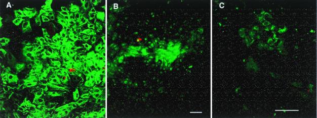
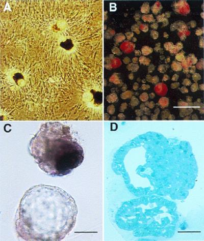
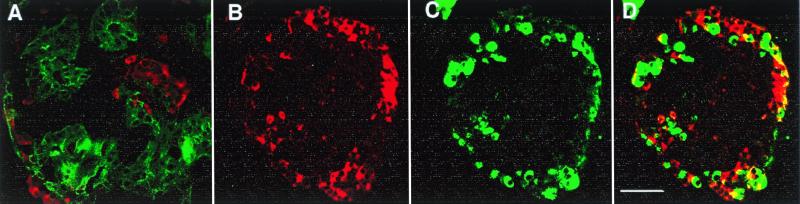
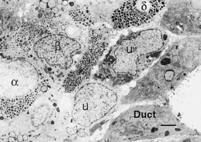
A major obstacle to successful islet transplantation for both type 1 and 2 diabetes is an inadequate supply of insulin-producing tissue. This need for transplantable human islets has stimulated efforts to expand existing pancreatic islets and/or grow new ones. To test the hypothesis that human adult duct tissue could be expanded and differentiated in vitro to form islet cells, digested pancreatic tissue that is normally discarded from eight human islet isolations was cultured under conditions that allowed expansion of the ductal cells as a monolayer whereupon the cells were overlaid with a thin layer of Matrigel. With this manipulation, the monolayer of epithelial cells formed three-dimensional structures of ductal cysts from which 50-to 150- micrometer diameter islet-like clusters of pancreatic endocrine cells budded. Over 3-4 weeks culture the insulin content per flask increased 10- to 15-fold as the DNA content increased up to 7-fold. The cultivated human islet buds were shown by immunofluorescence to consist of cytokeratin 19-positive duct cells and hormone-positive islet cells. Double staining of insulin and non-beta cell hormones in occasional cells indicated immature cells still in the process of differentiation. Insulin secretion studies were done over 24 h in culture. Compared with their basal secretion at 5 mM glucose, cysts/cultivated human islet buds exposed to stimulatory 20 mM glucose had a 2.3-fold increase in secreted insulin. Thus, duct tissue from human pancreas can be expanded in culture and then be directed to differentiate into glucose responsive islet tissue in vitro. This approach may provide a potential new source of pancreatic islet cells for transplantation.


Figures

Figure 1
Figure 1
(A) The adherent cells are primarily epithelial cells, immunostained for pan-cytokeratin (FITC green); staining for pan-cytokeratin and cytokeratin 19 were similar. Insulin-positive cells (Texas red) are scattered and infrequent. (B and C) Double staining of insulin (red) and transcription factor IPF-1 (FITC, green). Besides insulin-producing β cells, many duct cells express this transcription factor, both in the nucleus and in the cytoplasm. In B a number of cells express IPF-1 in the nucleus and/or cytoplasm without insulin staining; the field has the same density of cells as A. In addition, as in C, scattered clumps of cells had cytoplasmic IPF-1 staining with little nuclear staining and no insulin staining. Both A and B are 7-day cultured tissue of pancreas H99–12 pellet, whereas C is 7-day cultured tissue of pancreas H99–10 middle layer. (Magnification bars = 50 μm.)
Figure 2
Figure 2
(A) After ducts were overlaid with matrix, three-dimensional structures of ductal cysts with protruding buds of islet tissue (CHIBs) were observed rising from the monolayer lawn of cells. (B and C) There are variable numbers of dithizone-stained β cells in these harvested cycts/CHIBs; many of the structures are solely cysts whereas other have 50- to 150-μm islet buds. (D) The structure of budding islet cells from a cyst is seen in this toluidine blue 1-μm section. (Magnification bar = 500 μm in B, 100 μm in C, and 50 μm in D.)
Figure 3
Figure 3
Double immunostained sections of CHIBs. (A) Cytokeratin 19 (FITC, green)-positive duct cells make up most of this CHIB with insulin-positive cells (Texas red) in several islet buds. Another CHIB shown with cell positive for insulin (red in B and D) and for the non-β cell hormones (glucagon, somatostatin, and pancreatic polypeptide) (green in C and D); D is the overlay of these red and green images. There are a few cells that coexpress both β and non-β cell hormones (yellow in D), indicating that some of the cells are immature and still in differentiation. (Magnification bar = 50 μm.)
Figure 4
Figure 4
By ultrastructural analysis of CHIBs, mature and immature phenotypes could be seen. The duct cells (D), with characteristic short stubby microville and apical junctional complexes, line the lumen of a cyst. Adjacent to the ductal epithelium is a row of unidentifiable cells (U) that do not have characteristic granules of islet endocrine cells. β, α, and δ cells are identified by their granules. (Magnification bar = 2 μm.)
Figure 5
Figure 5
The cysts/CHIBs are responsive to glucose in vitro secretion studies. Forty cysts/CHIBs were incubated in six replicates from each of two groups for three pancreases. After an initial 4-h preincubation, each sample was incubated for 24 h in RPMI with 5 mM glucose for basal secretion determination. The media were replaced with either fresh media with 5 mM (hatched bars) or 20 mM (solid bars) glucose for an additional 24 h. Insulin secretion over the 24-h period was expressed as percent of the same tube at basal. Parallel experiments with middle layer tissue from two flasks of pancreas H99–19 and of H99–25 and one flask from H99–24 are shown. Insulin content was not determined.

References

    1. Weir G C, Bonner-Weir S. Diabetes. 1997;46:1247–1256. - PubMed
    1. Hering B J, Ricordi C. Graft. 1999;2:12–27.
    1. Brelje T C, Scharp D W, Lacy P E, Ogren L, Talamantes F, Robertson M, Friesen H G, Sorenson R L. Endocrinology. 1993;132:879–887. - PubMed
    1. Beattie G M, Cirulli V, Lopez A D, Hayek A. J Clin Endocrinol Metab. 1997;82:1852–1856. - PubMed
    1. Beattie G M, Itkin-Ansari P, Cirulli V, Leibowitz G, Lopez A D, Bossie S, Mally M I, Levine F, Hayek A. Diabetes. 1999;48:1013–1019. - PubMed

Publication types

LinkOut - more resources