. 2000 Jul 5;97(14):8081-6.
doi: 10.1073/pnas.97.14.8081.

Experience-dependent plasticity in the auditory cortex and the inferior colliculus of bats: role of the corticofugal system

Affiliations

Experience-dependent plasticity in the auditory cortex and the inferior colliculus of bats: role of the corticofugal system

E Gao et al. Proc Natl Acad Sci U S A. .

Abstract

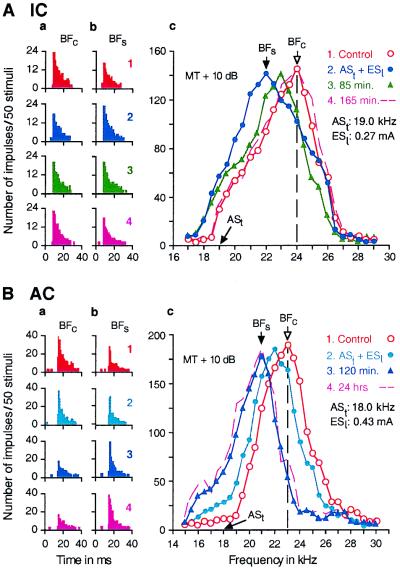
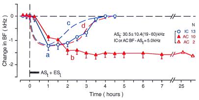
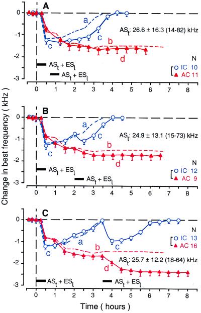
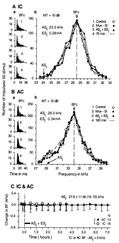
In the big brown bat, Eptesicus fuscus, the response properties of neurons and the cochleotopic (frequency) maps in the auditory cortex (AC) and inferior colliculus can be changed by auditory conditioning, weak focal electric stimulation of the AC, or repetitive delivery of weak, short tone bursts. The corticofugal system plays an important role in information processing and plasticity in the auditory system. Our present findings are as follows. In the AC, best frequency (BF) shifts, i.e., reorganization of a frequency map, slowly develop and reach a plateau approximately 180 min after conditioning with tone bursts and electric-leg stimulation. The plateau lasts more than 26 h. In the inferior colliculus, on the other hand, BF shifts rapidly develop and become the largest at the end of a 30-min-long conditioning session. The shifted BFs return (i. e., recover) to normal in approximately 180 min. The collicular BF shifts are not a consequence of the cortical BF shifts. Instead, they lead the cortical BF shifts. The collicular BF shifts evoked by conditioning are very similar to the collicular and cortical BF shifts evoked by cortical electrical stimulation. Therefore, our working hypothesis is that, during conditioning, the corticofugal system evokes subcortical BF shifts, which in turn boost cortical BF shifts. The cortical BF shifts otherwise would be very small. However, whether the cortical BF shifts are consequently boosted depends on nonauditory systems, including nonauditory sensory cortices, amygdala, basal forebrain, etc., which determine the behavioral relevance of acoustic stimuli.


Figures

Figure 1
Figure 1
Changes in the responses (a and b) and frequency-response curves (c) of a single collicular neuron (A) and a single cortical neuron (B) evoked by a 30-min-long conditioning session consisting of 60 stimulus pairs of a short ASt followed by an ESl. All of the data were obtained with tone bursts fixed at 10 dB above the minimum threshold (MT) of a given neuron. In A, ASt was 19.0 kHz, and the BF of the collicular neuron was 24.0 kHz. In B, ASt was 18.0 kHz, and the BF of the cortical neuron was 23.0 kHz. The data in A and B were obtained before (1, control condition), immediately after (2), 85 or 120 min after (3), and 165 min or 24 h after conditioning (4). The poststimulus time histograms in columns a and b, respectively, show the changes in the responses at the BFs in the control (BFc) and “shifted” conditions (BFs), which are indicated by arrows in c. The BF shift of the collicular neuron recovered (i.e., BFs shifted back to BFc) 165 min after the conditioning, but that of the cortical neuron did not recover even 24 h after the conditioning.
Figure 2
Figure 2
Time courses of the BF shifts of collicular (curve a, open circles) and cortical neurons (curve b, filled triangles) evoked by a 30-min-long conditioning session consisting of ASt + ESl (horizontal bar). The time course is quite different between the collicular and cortical neurons. Each data point represents the mean and standard error of the values obtained from the number of neurons (N) listed on the right. The mean frequency of ASts ranged from 19 to 60 kHz (30.5 ± 10.4 kHz). The BFs of 13 collicular and 10 cortical neurons studied were 5.0 kHz higher than the frequency of ASt. For comparison, the time courses of the BF shifts of collicular and cortical neurons evoked by a focal electrical stimulation of the AC are also shown in the figure by the dashed curves c and d, respectively (based on refs. and 7). These two dashed curves are similar to curve a.
Figure 3
Figure 3
Time courses of the BF shifts of collicular (curve c, open circles) and cortical neurons (curve d, filled triangles) evoked by two conditioning sessions with an identical 30-min-long conditioning session consisting of ASt + ESl (horizontal bars). The time interval between the first and second conditioning sessions was 10 (A), 90 (B), or 180 min (C). Each data point represents the mean and standard error of the values obtained from the number of neurons (N) listed on the right. The mean frequency and the range of ASts are listed in each figure. The BFs of the neurons studied were 5.0 kHz higher than the frequency of ASt. The dashed curves a and b represent the time courses of collicular (curve a) and cortical BF shifts (curve b) evoked by the first conditioning session alone and are the same as curves a and b in Fig. 2.
Figure 4
Figure 4
Bilateral inactivation of the somatosensory cortex with 0.4 μg of muscimol abolished changes in the responses (a) and frequency-response curves (b) of a single collicular neuron (A) and a single cortical neuron (B) that otherwise would be evoked by auditory conditioning. The poststimulus time histograms in a display the responses to tone bursts at the BF of a given neuron before (1, control condition); during muscimol application to the somatosensory cortices (2); immediately after conditioning under bilateral inactivation (3); and 75 or 180 min after conditioning (4). The frequency-response curves in b were obtained in the above four conditions. The frequencies of conditioned ASt and the current of unconditioned ESl are listed in b. BFc, BF in the control condition. (C) No changes in collicular (filled circles) and cortical BFs (filled triangles) were evoked by the conditioning with ASt + ESl (horizontal bar) because of bilateral inactivation of the somatosensory cortex. Each data point represents the mean and standard error of the values obtained from 16 collicular or 13 cortical neurons (as indicated on the right, N). The mean frequency and the range of ASts used were 27.8 ± 11.56 kHz and 19–72 kHz, respectively. The BFs of 29 neurons studied were 5.0 kHz higher than the frequency of ASt. All of the data were obtained with a tone burst delivered at 10 dB above the minimum threshold (MT) of a given neuron.

References

    1. Irvine D R, Rajan R. Clin Exp Pharmacol Physiol. 1996;23:939–947. - PubMed
    1. Weinberger N M. Neurobiol Learn Mem. 1998;70:226–251. - PubMed
    1. Buonomano D V, Merzenich M M. Annu Rev Neurosci. 1998;21:149–186. - PubMed
    1. Gao E, Suga N. Proc Natl Acad Sci USA. 1998;95:12663–12670. - PMC - PubMed
    1. Yan W, Suga N. Nat Neurosci. 1998;1:54–58. - PubMed

Publication types

LinkOut - more resources