

Epstein-Barr virus (EBV) in infectious mononucleosis: detection of the virus in tonsillar B lymphocytes but not in desquamated oropharyngeal epithelial cells
- PMID: 10884920
- PMCID: PMC1186900
- DOI: 10.1136/mp.53.1.37
Epstein-Barr virus (EBV) in infectious mononucleosis: detection of the virus in tonsillar B lymphocytes but not in desquamated oropharyngeal epithelial cells
Abstract
Aims: Despite its well established tropism for B cells, the nature of the cellular compartment(s) mediating primary and persistent Epstein-Barr virus (EBV) infection is still a matter of controversy. In view of the association of EBV with several lymphoid and epithelial malignancies, resolution of this issue is important.
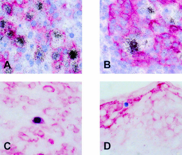
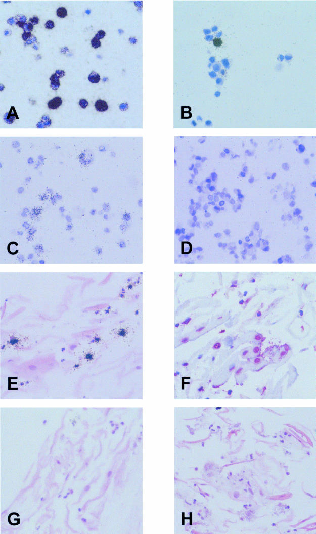
Methods: Desquamated oropharyngeal epithelial cells from 10 patients with acute infectious mononucleosis and from seven chronic virus carriers were studied for evidence of EBV infection using in situ hybridisation for the detection of the small EBV encoded RNAs (EBERs) and of the viral genome. In addition, immunocytochemistry was used to detect the BZLF1 transactivator protein of EBV.
Results: There was no evidence of latent or replicative EBV infection in oropharyngeal epithelial cells in any of the samples. In contrast, EBV infected B cells were readily identified in a tonsil from a patient with infectious mononucleosis.
Conclusions: The results suggest that oropharyngeal epithelial cells are not a major site of EBV infection and provide further support for the notion that B cells mediate primary and persistent EBV infection.
Figures
Similar articles
-
Morphology, immunophenotype, and distribution of latently and/or productively Epstein-Barr virus-infected cells in acute infectious mononucleosis: implications for the interindividual infection route of Epstein-Barr virus.Blood. 1995 Feb 1;85(3):744-50. Blood. 1995. PMID: 7530505
-
Epstein-Barr virus (EBV) infection in infectious mononucleosis: virus latency, replication and phenotype of EBV-infected cells.J Pathol. 1997 Jun;182(2):151-9. doi: 10.1002/(SICI)1096-9896(199706)182:2<151::AID-PATH824>3.0.CO;2-3. J Pathol. 1997. PMID: 9274524
-
Strict lymphotropism of Epstein-Barr virus during acute infectious mononucleosis in nonimmunocompromised individuals.Blood. 1997 Apr 15;89(8):2856-62. Blood. 1997. PMID: 9108405
-
Infectious mononucleosis and Epstein-Barr virus.Expert Rev Mol Med. 2004 Nov 5;6(23):1-16. doi: 10.1017/S1462399404008440. Expert Rev Mol Med. 2004. PMID: 15541197 Review.
-
[The entry of Epstein-Barr virus into B lymphocytes and epithelial cells during infection].Bing Du Xue Bao. 2014 Jul;30(4):476-82. Bing Du Xue Bao. 2014. PMID: 25272606 Review. Chinese.
Cited by
-
Epstein-Barr virus infection in the pathogenesis of nasopharyngeal carcinoma.Mol Pathol. 2000 Oct;53(5):248-54. doi: 10.1136/mp.53.5.248. Mol Pathol. 2000. PMID: 11091848 Free PMC article. Review.
-
Cancer stem-like cell: a novel target for nasopharyngeal carcinoma therapy.Stem Cell Res Ther. 2014;5(2):44. doi: 10.1186/scrt433. Stem Cell Res Ther. 2014. PMID: 25158069 Free PMC article. Review.
-
Identification of B cells as a major site for cyprinid herpesvirus 3 latency.J Virol. 2014 Aug;88(16):9297-309. doi: 10.1128/JVI.00990-14. Epub 2014 Jun 4. J Virol. 2014. PMID: 24899202 Free PMC article.
-
Molecular features and translational outlook for Epstein-Barr virus-associated gastric cancer.Future Virol. 2018 Nov;13(11):803-818. doi: 10.2217/fvl-2018-0071. Epub 2018 Oct 24. Future Virol. 2018. PMID: 34367314 Free PMC article.
-
Themed issue: the biology and pathology of the Epstein-Barr virus.Mol Pathol. 2000 Oct;53(5):219-21. doi: 10.1136/mp.53.5.219. Mol Pathol. 2000. PMID: 11091845 Free PMC article. No abstract available.
References
-
- Rickinson AB, Kieff E. Epstein-Barr virus. In: Fields BN, Knipe DM, Howley PM, eds. Fields virology, 2nd ed. Philadelphia: Lipincott-Raven, 1996:2397–446.
-
- Niedobitek G, Agathanggelou A, Nicholls JM. Epstein-Barr virus infection and the pathogenesis of nasopharyngeal carcinoma: viral gene expression, tumour cell phenotype, and the role of the lymphoid stroma. Semin Cancer Biol 1996;7:165–74. - PubMed
-
- Miller G. The switch between latency and replication of Epstein-Barr virus. J Infect Dis 1990;161:833–44. - PubMed
-
- Evans AS. Clinical syndromes associated with EB virus infection. Ann Intern Med 1972;18:77–93. - PubMed
Publication types
MeSH terms
Substances
LinkOut - more resources
Full Text Sources