Review
. 2000 Jul;13(3):470-511.
doi: 10.1128/CMR.13.3.470.

Pathogenesis of group A streptococcal infections

Affiliations
Review

Pathogenesis of group A streptococcal infections

M W Cunningham. Clin Microbiol Rev. 2000 Jul.

Abstract

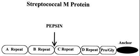
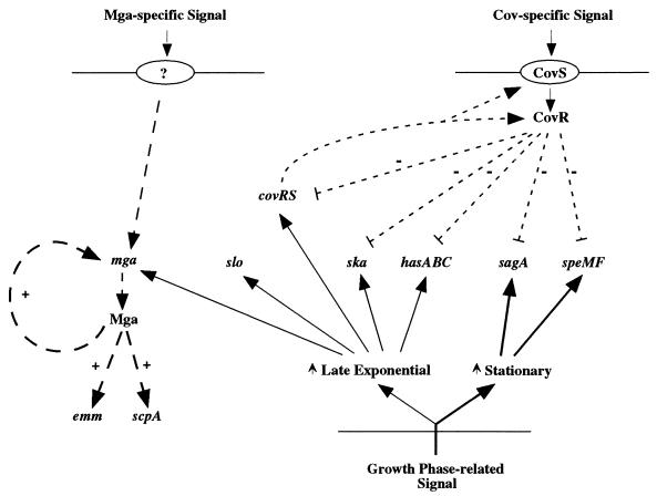
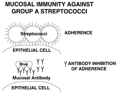
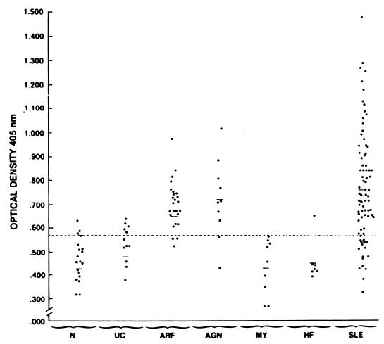
Group A streptococci are model extracellular gram-positive pathogens responsible for pharyngitis, impetigo, rheumatic fever, and acute glomerulonephritis. A resurgence of invasive streptococcal diseases and rheumatic fever has appeared in outbreaks over the past 10 years, with a predominant M1 serotype as well as others identified with the outbreaks. emm (M protein) gene sequencing has changed serotyping, and new virulence genes and new virulence regulatory networks have been defined. The emm gene superfamily has expanded to include antiphagocytic molecules and immunoglobulin-binding proteins with common structural features. At least nine superantigens have been characterized, all of which may contribute to toxic streptococcal syndrome. An emerging theme is the dichotomy between skin and throat strains in their epidemiology and genetic makeup. Eleven adhesins have been reported, and surface plasmin-binding proteins have been defined. The strong resistance of the group A streptococcus to phagocytosis is related to factor H and fibrinogen binding by M protein and to disarming complement component C5a by the C5a peptidase. Molecular mimicry appears to play a role in autoimmune mechanisms involved in rheumatic fever, while nephritis strain-associated proteins may lead to immune-mediated acute glomerulonephritis. Vaccine strategies have focused on recombinant M protein and C5a peptidase vaccines, and mucosal vaccine delivery systems are under investigation.


Figures

FIG. 1
FIG. 1
Electron micrographs demonstrating the attachment and internalization of streptococci by human cultured pharyngeal cells. Group A streptococci were observed to associate with microvilli upon initial contact with the pharyngeal cells. Membrane extension occurred during the internalization process. Surface interaction can be seen between the pharyngeal cell and streptococcus. Intracellular streptococci were found ingulfed in cytoplasmic vacuoles. Magnifications: A, ×12,700; B, ×24,300. (Reprinted from reference with permission from the publisher.)
FIG. 2
FIG. 2
How the immune system recognizes group A streptococci and uses opsonization by complement and type-specific antibody against M protein or any other surface molecule capable of generating opsonic antibody. Fc receptors shown on macrophages bind to the antibody Fc region, inducing phagocytosis and killing of the streptococci.
FIG. 3
FIG. 3
Diagram of the group A streptococcal cell covered with an outer hyaluronic acid capsule and the group A carbohydrate, consisting of a polymer of rhamnose with N-acetylglucosamine side chains. Streptococcal M protein extends from the cell wall and is anchored in the membrane. (Reprinted from reference with permission from the publisher.)
FIG. 4
FIG. 4
Electron micrograph of group A M type 24 streptococci. The surface fimbriae or hairlike projections indicate the presence of M protein on the surface of the streptococci.
FIG. 5
FIG. 5
The A, B, C, and D repeat regions of M protein, with the protein anchor and pepsin cleavage site shown. The A repeat region varies between serotypes and contains the highly variable serotype-specific amino acid sequence of M protein at the N terminus. The B repeat region varies from serotype to serotype, while the C repeat region contains a conserved sequence shared among all of the serotypes. The anchor region contains the LPXTGX motif required to anchor gram-positive proteins in the cell membrane. (Reprinted from reference with permission from the publisher.)
FIG. 6
FIG. 6
Overview of the regulatory networks in expression of group A streptococcal virulence genes. +, positive regulation; —, negative regulation. Short dashed lines indicate CovR regulation, and long dashed lines represent regulation by Mga. Solid lines show the correlation between growth phase (exponential [light lines] or stationary [darker lines]). As shown in the figure, Mga activates transcription of several genes, including those for M protein (emm), C5a peptidase (scpA), M-like proteins (mrp, enn, and fcR), serum opacity factor (sof), and secreted inhibitor of complement (sic) (95, 101, 355, 358, 422). Mga feeds back to positively regulate itself and functions as a 62-kDa protein to bind to the promotor region of genes that it regulates (417). The genes involved in the mga regulon are shown in Table 3, and an overview of regulation is shown in the diagram above. A global negative regulator of mga was identified in a few strains and called nra. In addition to repressing Mga synthesis 4- to 16-fold, nra was a negative regulator of prtF2, the gene for the fibronectin-binding protein F2, and the collagen-binding protein gene (cpa) in group A streptococci. The Nra protein sequence was 62% homologous to RofA, a positive transcriptional regulator of the fibronectin-binding protein (prtF) and itself in response to increased oxygen levels (426). RofA and Mga may influence the expression of genes involved in adhesion in different environments (426). csrR and csrS or covR and covS are a pair of genetic loci in group A streptococci that encode a two-component regulatory system (327). Inactivation of csrR or covR resulted in a striking increase in transcription of the capsule synthesis genes of the has operon and a corresponding increase in hyaluronic acid capsule production (327). Subsequent work confirmed these observations and demonstrated binding of the CsrR protein to the promoter region upstream of the has operon (42). The CsrR or CovR protein acts as a transcriptional regulator. Production of a nonpolar mutation in csrR or covR increased transcription of several other virulence genes, including ska (streptokinase), sagA (streptolysin O), and speF (mitogenic factor) but had no effect on mga, emm, scpA, speB, or speC. Thus, the CovR or CsrR response regulator repressed transcription of several virulence operons in group A streptococci (172). Since multiple unrelated genes were controlled by csrR, an alternative nomenclature was given to the csrR-csrS locus, covR-covS for control of virulence genes. The csrR-csrS or covR-covS sensor-regulator gene pair represent a new regulatory pathway affecting expression of several group A streptococcal virulence genes which are not regulated by mga and has been found in all group A streptococcal strains tested (172). (Reprinted from reference with permission from the publisher.)
FIG. 7
FIG. 7
Streptococcal adherence and inhibition of adherence to the mucosa by specific antibody. Mucosal antibody against surface adhesins or epitopes in the C repeat region of M proteins protects against colonization with group A streptococci.
FIG. 8
FIG. 8
Identification of an idiotype (My1) present on antibodies in sera from patients with acute rheumatic fever (ARF) and acute glomerulonephritis (AGN) as well as systemic lupus erythematosus (SLE) and Sjögren's syndrome (not shown). Sera from normal individuals (N) and patients with uncomplicated streptococcal disease (UC), heart failure (HF), Chagas' disease (not shown), and IgA nephropathy (not shown) are compared in the figure, and all have normal levels of the My1 idiotype. (Reprinted from reference with permission from the publisher.)
FIG. 9
FIG. 9
Reactivity of antistreptococcal-antimyosin MAb with human myocardium in an immunofluorescence assay. (Reprinted from reference with permission from the publisher. Copyright 1989. The American Association of Immunologists.)
FIG. 10
FIG. 10
Reactivity of antistreptococcal-antimyosin MAb 36.2.2 with the surface or extracellular matrix of rat myocardial cells in culture. MAb 36.2.2 exhibits cytotoxicity against rat heart cells in the presence of complement. (Reprinted from reference with permission from the publisher. Copyright 1997. The American Association of Immunologists.)
FIG. 11
FIG. 11
(A) Homology between human cardiac myosin residues 1313 to 1329 and streptococcal M5 protein peptide B2 (M5 residues 150 to 167). Identity (47%) was observed in a 17-amino-acid overlap. The M5 peptides B2, B1B2, and B3A contain large amounts of overlapping sequence. :, identical residues; ., conserved substitutions. Three of the residues shown in panel A in the myosin sequence are unique to human cardiac myosin. (B) Amino acid sequence identity (LKTEN) between M5 peptide NT4 and human cardiac myosin (LQTEN). NT4 contains residues 40 to 58 of the M5 protein. The cardiac myosin sequence shown is found in residues 1279 to 1286 near the beginning of the light meromyosin tail and the end of the S-2 fragment of myosin. The myosin sequence shown in panel B is conserved among cardiac myosins. (C) Amino acid sequence identity between serotype M6 protein and human cardiac myosin within the same regions. The repeat in the M6 protein is LTTEN, which is repeated five times in the N-terminal region of the M6 protein. In M5 and M6 proteins, the LKTEN and LTTEN sequences, respectively, are conserved and are similar to the LQTEN sequence in cardiac myosins. (Reprinted from reference with permission from the publisher.)

References

    1. Adderson E E, Shikhman A R, Ward K E, Cunningham M W. Molecular analysis of polyreactive monoclonal antibodies from rheumatic carditis: human anti-N-acetyl-glucosamine/anti-myosin antibody V region genes. J Immunol. 1998;161:2020–2031. - PubMed
    1. Adrezejewski C, Jr, Rauch J, Stollar B D, Schwartz R S. Antigen binding diversity and idiotypic cross-reactions among hybridoma autoantibodies to DNA. J Immunol. 1980;126:226–231. - PubMed
    1. Ahmed S, Ayoub E M. Severe, invasive group A streptococcal disease and toxic shock. Pediatr Ann. 1998;27:287–292. - PubMed
    1. Ahmed S, Ayoub E M, Scornik J C, Wang C-Y, She J-X. Poststreptococcal reactive arthritis: clinical characteristics and association with HLA-DR alleles. Arthritis Rheum. 1998;41:1096–1102. - PubMed
    1. Akerstrom B, Brodin T, Reis K, Bjorck L. Protein G: a powerful tool for binding and detection of monoclonal and polyclonal antibodies. J Immunol. 1985;135:2589–2592. - PubMed

Publication types

MeSH terms

LinkOut - more resources