. 2000 Jul 1;14(13):1617-30.

Conditional biallelic Nf2 mutation in the mouse promotes manifestations of human neurofibromatosis type 2

Affiliations

Conditional biallelic Nf2 mutation in the mouse promotes manifestations of human neurofibromatosis type 2

M Giovannini et al. Genes Dev. .

Abstract

Hemizygosity for the NF2 gene in humans causes a syndromic susceptibility to schwannoma development. However, Nf2 hemizygous mice do not develop schwannomas but mainly osteosarcomas. In the tumors of both species, the second Nf2 allele is inactivated. We report that conditional homozygous Nf2 knockout mice with Cre-mediated excision of Nf2 exon 2 in Schwann cells showed characteristics of neurofibromatosis type 2. These included schwannomas, Schwann cell hyperplasia, cataract, and osseous metaplasia. Thus, the tumor suppressor function of Nf2, here revealed in murine Schwann cells, was concealed in hemizygous Nf2 mice because of insufficient rate of second allele inactivation in this cell compartment. The finding of this conserved function documents the relevance of the present approach to model the human disease.


Figures

Figure 1
Figure 1
Generation and functional analysis of the Nf2KO3, Nf2Δ2, and Nf2flox2 alleles. (A) Targeting strategies. Restriction maps of the Nf2+ allele (5′ portion), the Nf2KO3 and the Nf2lox2 targeting fragments, and the different modified Nf2 alleles after homologous recombination and Cre-mediated recombination. Exons 2, 3, and 4 are indicated (black boxes) and restriction sites used for cloning and screening (A) ApaI, (B) BamHI, (Bs) BstBI, (E) EcoRI, (EV) EcoRV, (K) KpnI, (P) PstI, (S) SacI, (Sp) SpeI, (X) XbaI, (Xh) XhoI. The Nf2KO3 targeting fragment comprises the IRESLacZPGKHyg selection cassette, inserted into the BstBI site of exon 3 in the Nf2 orientation. The Nf2lox2 targeting fragment comprises the floxed PGKHprt selection cassette, inserted into the BamHI site downstream of Nf2 exon 2 in opposite orientation to Nf2, plus a third loxP site that was introduced into the EcoRI site upstream of exon 2. All loxP sites are represented by open triangles. Recombination of loxP2 and loxP3 results in the loxP2+3 site, and the loxP1+3 is the result of recombination of loxP1 and loxP3. In the first step of the strategy, the Nf2 locus was targeted with the Nf2lox2 targeting fragment. In the second step, the PGKHprt selection cassette was removed by Cre recombinase that, upon transient expression in Nf2lox2/+ ES cells, recombined loxP1+3 and loxP2+3 sites (ES cell clones with recombined loxP1+2 sites are lost by 6-TG selection). The double-headed arrows indicate the DNA fragments resulting from digestions with different enzymes expected to hybridize with probes A or B. Also depicted are combinations of PCR primers P1–6 that detect the different Nf2 alleles. (B) Mutant schwannomins produced by the Nf2KO3 and Nf2Δ2 alleles in heterozygous mutant mice are present at strongly reduced amounts compared to wild-type schwannomin. Immunoprecipitations and immunoblotting were performed on brain extracts of wild-type and mutant Nf2 mice using two different anti-NF2 polyclonal antibodies. The bands correspond to the indicated proteins.
Figure 2
Figure 2
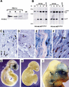
Cre expression and activity pattern in P0Cre transgenic mice. (A) Cre expression in sciatic nerves of adult P0CreA–C transgenic mice. Western blot analysis with rabbit polyclonal anti-Cre antibody. As indicated by a positive immunoreaction at 38 kD, the three transgenic lines show Cre expression in sciatic nerves. (B) Cre-mediated deletion in adult mouse tissues. DNA of various tissues of a P0CreB;Nf2flox2/flox2 and a P0CreC; Nf2flox2/flox2 mouse (both 3-month-old) was analyzed by Southern blotting (XbaI–BamHI digestion, probe B). The Nf2flox2 allele (5.0 kb) and Nf2Δ2 allele (3.0 kb) can be distinguished by a XbaI–BamHI digestion, but the Nf2flox2 and the Nf2+ alleles (both 5.0 kb) cannot. (T) Trigeminal nerve, (S) sciatic nerve, (B) brachial nerve, (Le) lens, (Li) liver, (Nf2Δ2/+) tail (1 : 1 ratio of Nf2+ and Nf2Δ2 allele). Arrows on the left side refer to the marker. (C,D) Cre-expressing cells in sciatic nerves of adult P0CreB (C) and P0CreC (D) transgenic mice, as detected by immunostaining with anti-Cre antibodies. Positive nuclei are indicated by arrows. (E,F) β-Galactosidase activity in sciatic nerves of adult P0CreA;floxlacZ double-transgenic mice, as detected by whole-mount X-gal staining and counterstaining of the sections with nuclear fast red. (E) Sagittal section. (F) Sagittal section additionally immunostained with anti-Krox-20 antibodies. A detail is shown of an area with homogeneously blue-stained cells. Recombination occurs in myelinated (Krox-20+, black arrow) as well as in non-myelinated (Krox-20, white arrow) Schwann cells. (G–I) Temporal and spatial expression of lacZ in whole-mount X-gal-stained P0Cre;floxlacZ double-transgenic mouse embryos. (G) P0CreA;floxlacZ embryo at E9.5. Lateral view demonstrating that β-galactosidase activity is mostly observed in the area of the neural crest cell migration: head mesenchyme, ventral cranio-facial area (hm), otocyst (ot), and facio-acoustic neural crest complex (7-8). (H) P0CreC;floxlacZ embryo at E9.5. Lateral view demonstrating scattered areas of weak β-galactosidase activity (arrows). (I) P0CreA;floxlacZ embryo at E12.5. Lateral view demonstrating β-galactosidase activity in the frontonasal region of the head mesenchyme (hm), ganglia of the VIIIth (VIII) and Vth (V) cranial nerves, and brachial plexus (bp). Magnification, (CF) 40×.
Figure 3
Figure 3
Survival of conditional Nf2 knockout mice and DNA analysis of tumors. (A) The viability of P0Cre;Nf2flox2/flox2 mice correlates with the expression level of Cre. Survival curves of P0CreA–D;Nf2flox2/flox2 and Nf2flox2/flox2 mice over a period of 24 months (numbers of considered moribund plus dead animals in brackets). (B) Tumors of P0Cre;Nf2flox2/flox2 mice show Cre-mediated Nf2 gene inactivation. Southern blot analysis (probe B) of XbaI–BamHI-digested DNA of nine representative tumors (lanes 19). The bands corresponding to the Nf2flox2 and Nf2Δ2 allele, and those referring to the marker (left) are indicated by arrows. (C) Tumors of P0Cre;Nf2KO3/flox2 mice show Nf2 gene inactivation. Southern blot analysis (probe B) of ApaI–SpeI-digested DNA of a neurofibrosarcoma in the uterus (lane 1) and the corresponding metastasis in a lymph node (lane 2). The primary tumor and the metastasis showed Cre-mediated excision of the floxed exon 2: The intensity of the Nf2Δ2 band inversely correlates with that of the Nf2flox2 band, indicating that no loss of the Nf2Δ2 allele has occurred. All different Nf2 alleles can be distinguished by ApaI–SpeI digestion, as indicated in Fig. 1A (Nf2+ > 13 kb). (D) Osseous metaplasia in the lung of a P0Cre;Nf2flox2/flox2 mouse shows Cre-mediated Nf2 gene inactivation. Southern blot analysis (probe B) of XbaI–BamHI-digested DNA obtained from the lesion shown in Fig. 5C (lane 1). The bands corresponding to the Nf2flox2 and Nf2Δ2 allele are indicated by arrows.
Figure 4
Figure 4
Histological analysis of phenotypic abnormalities in P0Cre;Nf2flox2/flox2 mice. (A,B) Schwann cell tumors. (A) P0CreC line. Spindle cell tumor located on the external side of the esophageal-gastric junction. The tumor shows focal Schwann cell characteristics such as Antoni A type palisading (arrows) and primitive Verocay body formation. (E) P0CreC line. Ultrastructural examination of a uterine schwannoma showed Schwann cells with long, thin cytoplasmic processes (arrow) and variantly coated by a basement membrane (BM). Schwann cell nucleus (N). (C–I) Schwann cell hyperplasia. (C,D) P0CreB line. (C) Schwann cell hyperplasia (arrows) in a trigeminal ganglion and (D) normal trigeminal ganglion of a wild-type littermate. (E) P0CreA line. Schwann cell hyperplasia/atrophy (arrows) in a sciatic nerve. (F–I) Ultrastructure of the sciatic nerves of a (F,H) Nf2flox2/flox2 and (G,I) P0CreA;Nf2flox2/flox2 mouse. (F,H) Normally myelinated Schwann cell (M) surrounding the axoplasm (Ax). Non-myelinated Schwann cells (NM) are also shown. (G,I) Myelin sheaths herniating and looping into the central axonal region of the nerve. (A,C–E) H&E stains. Magnification in (A) 200×; (B) 4400×; (C–E) 630×; (F) 3000×; (G) 4400×; (H) 12000×; (I) 10,400×.
Figure 5
Figure 5
P0-specific conditional homozygous Nf2 knockout mice develop characteristics of neurocristopathy. (A) Osteoma developing within the main mandibular nerve. (B) Odontoma of upper incisor, deforming the maxilla and nasal septum. Many foci of “ectopic” enamel production are seen (arrow) as well as osteopetrotic processes in the contralateral maxilla. (C) Osseous metaplasia (arrow) in the lung. (D) Cataract in a mouse showing normal cranio-facial development. Note the small, irregularly shaped lens (L) and the attachment of the lens to the cornea (Co). Retina (R). H&E stains. Magnification, (A) 400×; (B,D) 25×; (C) 100×.

References

    1. Akagi K, Sandig V, Vooijs M, Van der Valk M, Giovannini M, Strauss M, Berns A. Cre-mediated somatic site-specific recombination in mice. Nucleic Acids Res. 1997;25:1766–1773. - PMC - PubMed
    1. Bianchi AB, Mitsunaga S-I, Cheng JQ, Klein WM, Jhanwar SC, Seizinger B, Kley N, Klein-Szanto AJP, Testa JR. High frequency of inactivating mutations in the neurofibromatosis type 2 gene (NF2) in primary malignant mesotheliomas. Proc Natl Acad Sci. 1995;92:10854–10858. - PMC - PubMed
    1. Bolande R. The neurochristopathies. A unifying concept of disease arising in neural crest maldevelopment. Hum Pathol. 1974;5:409–420. - PubMed
    1. Couly GF, Coltey PM, Le Douarin NM. The triple origin of skull in higher vertebrates: A study in quail-chick chimeras. Development. 1993;117:409–429. - PubMed
    1. Deguen B, Merel P, Goutebroze L, Giovannini M, Reggio H, Arpin M, Thomas G. Impaired interaction of naturally occurring mutant NF2 protein with actin-based cytoskeleton and membrane. Hum Mol Genet. 1998;7:217–226. - PubMed

Publication types