. 2000 Aug;74(15):6725-33.
doi: 10.1128/jvi.74.15.6725-6733.2000.

Effects of palmitoylation of replicase protein nsP1 on alphavirus infection

Affiliations

Effects of palmitoylation of replicase protein nsP1 on alphavirus infection

T Ahola et al. J Virol. 2000 Aug.

Abstract

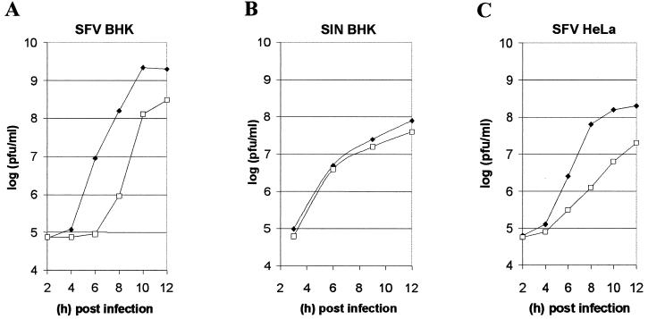
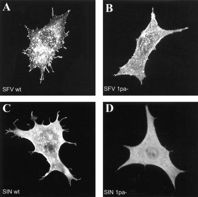
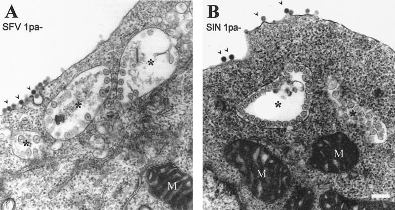
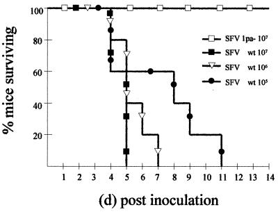
The membrane-associated alphavirus RNA replication complex contains four virus-encoded subunits, the nonstructural proteins nsP1 to nsP4. Semliki Forest virus (SFV) nsP1 is hydrophobically modified by palmitoylation of cysteines 418 to 420. Here we show that Sindbis virus nsP1 is also palmitoylated on the same site (cysteine 420). When mutations preventing nsP1 palmitoylation were introduced into the genomes of these two alphaviruses, the mutant viruses remained viable and replicated to high titers, although their growth was slightly delayed. The subcellular distribution of palmitoylation-defective nsP1 was altered in the mutant: it no longer localized to filopodial extensions, and a fraction of it was soluble. The ultrastructure of the alphavirus replication sites appeared normal, and the localization of the other nonstructural proteins was unaltered in the mutants. In both wild-type- and mutant-virus-infected cells, SFV nsP3 and nsP4 could be extracted from membranes only by alkaline solutions whereas the nsP2-membrane association was looser. Thus, the membrane binding properties of the alphavirus RNA replication complex were not determined by the palmitoylation of nsP1. The nsP1 palmitoylation-defective alphaviruses produced normal plaques in several cell types, but failed to give rise to plaques in HeLa cells, although they induced normal apoptosis of these cells. The SFV mutant was apathogenic in mice: it caused blood viremia, but no infectious virus was detected in the brain.


Figures

FIG. 1
FIG. 1
Palmitoylation of wt and mutant forms of SFV and SIN nsP1. (A) Amino acid sequence comparison of representative alphaviruses (36) in the vicinity of the palmitoylated cysteines of nsP1. Cysteine residues are shown in boldface type, and the number of the first amino acid is indicated. Arrows denote the mutation(s) from cysteine to alanine constructed to prevent the palmitoylation of nsP1 in SFV1pa− and SIN1pa−. RRV, Ross River virus; ONN, O'Nyong-Nyong virus; VEE, Venezuelan equine encephalitis virus. (B) Palmitoylation of nsP1 was analyzed by labeling of cells expressing nsP1 alone (Transfected) or in the context of alphavirus infection (Infected) with [3H]palmitate. Mutant or wt forms of SFV or SIN nsP1 were used as indicated at the top. The cell extracts were immunoprecipitated with antisera against nsP1 and analyzed by SDS-polyacrylamide gel electrophoresis and fluorography to visualize radioactive labeling of nsP1. Equal amounts of each wt-mutant pair of samples were loaded on the gel. Approximately 10 times more infected cell extracts than transfected cell extracts were used in this analysis due to the higher level of expression of the proteins in transfected cells. (C) The labeled cell extracts used in panel B were analyzed by Western blotting with antiserum against nsP1. Equal amounts of each wt-mutant pair were loaded on the gel.
FIG. 2
FIG. 2
One-step growth curves of SIN and SFV. The wt (solid diamond) or nsP1 palmitoylation-defective (1pa−) (open square) SFV (A and C) and SIN (B) were used to infect BHK or HeLa cells, as indicated (MOI = 5). Duplicate samples were collected at the indicated time points, and the amount of infectious virus was measured by plaque formation in BHK cells. This experiment was done three times with closely similar results; results from a representative experiment are shown.
FIG. 3
FIG. 3
Localization of nsP1 in infected cells. BHK cells infected with SFVwt (A), SFV1pa− (B), SINwt (C), or SIN1pa− (D) were fixed at 5 h (A and B) or 6 h (C and D) postinfection and stained by indirect immunofluorescence using anti-nsP1 antibodies. Magnification, ×875.
FIG. 4
FIG. 4
Ultrastructure of the CPVIs in infected cells. BHK cells infected for 5 h with SFV1pa− (A) and SIN1pa− (B) were processed for transmission electron microscopy. The sections shown contain CPVIs (asterisks) with spherules at their surrounding membrane, as well as mitochondria (M), nucleus (N), and budding viruses (arrowheads) at the plasma membrane. Bar, 200 nm.
FIG. 5
FIG. 5
Membrane association of SFV nonstructural proteins. (A) BHK cells were infected with SFVwt or SFV1pa− (as indicated) for 3.5 h and then treated with cycloheximide (100 μg/ml) for 0.5 h to allow the maturation and intracellular transport of the proteins previously produced. The cells were cooled on ice, lysed, and fractionated to yield cytoplasmic membranes pelleting at 15,000 × g and the remaining supernatant. The nuclei were discarded. Equal amounts of the pellet (p) and supernatant (s) fractions were analyzed for the presence of each of the nonstructural proteins by Western blotting, as indicated. The position of nsP4 is indicated by an arrow, since additional nonspecific bands are frequently observed. (B) BHK cells were infected with SFVwt for 3.5 h or with SFV1pa− for 4 h, followed by a 0.5-h treatment with cycloheximide and preparation of P15 membranes as above. The membranes were treated for 30 min on ice with 50 mM Tris (pH 7.5)–100 mM NaCl (lanes TN), 50 mM Tris–1 M NaCl (lanes NaCl), 75 mM NaCO3 (pH 11.5) (lanes 11.5), or 75 mM NaCO3 (pH 12) (lanes 12), followed by reisolation of the membranes by centrifugation. Equal portions of the resulting membrane pellets and supernatants were analyzed by Western blotting for each of the nonstructural proteins, as indicated on the left.
FIG. 6
FIG. 6
Plaque morphology of SFVwt and SFV1pa−. (A to D) BHK cells (A and B) or HeLa cells (C and D) were infected with either SFVwt (A and C) or SFV1pa− (B and D). The cells were incubated under a CMC layer for 2 days to allow plaque formation and stained with crystal violet before being photographed. (E to H). HeLa cell plaques on day 2 after SFVwt infection (E and F) or on day 4 after SFV1pa− infection (G and H) were stained by indirect immunofluorescence using antibodies against nsP1 (E and G) and by the TUNEL stain (F and H) to detect apoptotic cells. The center and edge of a plaque are indicated. Magnification, ×3 (panels A through D) and ×35 (panels E through H).
FIG. 7
FIG. 7
Pathogenicity of SFVwt and SFV1pa− in mice. Five BALB/c mice were inoculated intraperitoneally with each indicated dose (PFU) of the two viruses. Mouse survival is plotted against the time after inoculation. The experiment was carried out so that the identity of the virus (wt or mutant) used in the infections was not known to the persons performing the experiment. This experiment was carried out twice with closely similar results; data from one experiment are shown.
FIG. 8
FIG. 8
SFVwt or SFV1pa− titers in infected mice. Samples were collected on each day after inoculation of mice with 106 PFU of the respective viruses, as described in Materials and Methods. Two mice were sacrificed each day for each virus. The amount of infectious virus in blood (A and B) and in brain tissue samples (C and D) was measured by a plaque assay in MBA-13 cells. The asterisks indicate living mice without detectable virus titer, and crosses indicate mice killed by the virus.

References

    1. Ahola T, Kääriäinen L. Reaction in alphavirus mRNA capping: formation of a covalent complex of nonstructural protein nsP1 with 7-methyl-GMP. Proc Natl Acad Sci USA. 1995;92:507–511. - PMC - PubMed
    1. Ahola T, Laakkonen P, Vihinen H, Kääriäinen L. Critical residues of Semliki Forest virus RNA capping enzyme involved in methyltransferase and guanylyltransferase-like activities. J Virol. 1997;71:392–397. - PMC - PubMed
    1. Ahola T, Lampio A, Auvinen P, Kääriäinen L. Semliki Forest virus RNA capping enzyme requires association with anionic membrane phospholipids for activity. EMBO J. 1999;18:3164–3172. - PMC - PubMed
    1. Atkins G J, Sheahan B J, Liljeström P. The molecular pathogenesis of Semliki Forest virus: a model virus made useful? J Gen Virol. 1999;80:2287–2297. - PubMed
    1. Barton D J, Sawicki S G, Sawicki D L. Solubilization and immunoprecipitation of alphavirus replication complexes. J Virol. 1991;65:1496–1506. - PMC - PubMed

Publication types

MeSH terms

LinkOut - more resources