. 2000 Aug;74(15):7137-45.
doi: 10.1128/jvi.74.15.7137-7145.2000.

Effects of truncation of the carboxy terminus of pseudorabies virus glycoprotein B on infectivity

Affiliations

Effects of truncation of the carboxy terminus of pseudorabies virus glycoprotein B on infectivity

R Nixdorf et al. J Virol. 2000 Aug.

Abstract

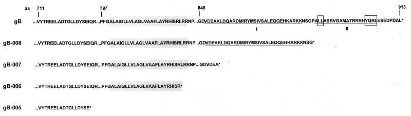
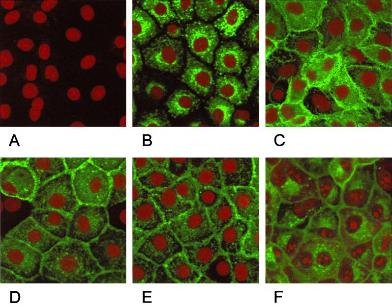
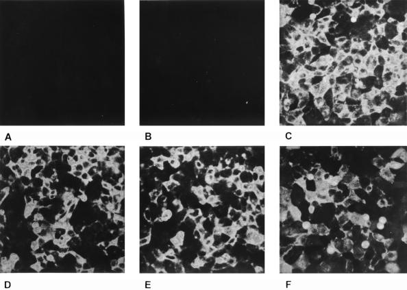
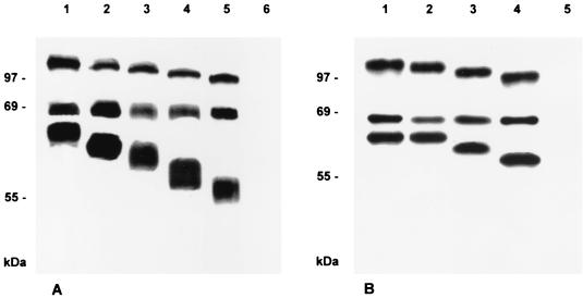
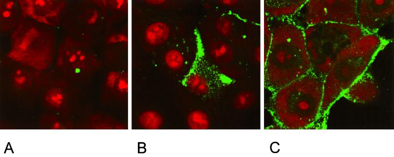
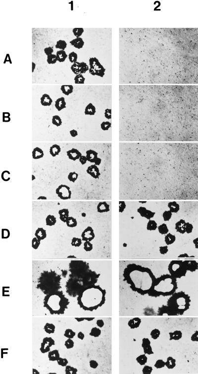
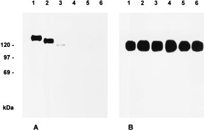
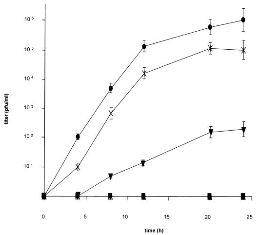
Glycoproteins homologous to the type I membrane glycoprotein B (gB) of herpes simplex virus 1 (HSV-1) are the most highly conserved glycoproteins within the family Herpesviridae and are present in members of each herpesvirus subfamily. In the alphaherpesvirus pseudorabies virus (PrV), gB is required for entry into target cells and for direct viral cell-to-cell spread. These processes, though related, appear to be distinct, and thus it was interesting to analyze whether they require different functions of gB. To this end, we established cell lines stably expressing different carboxy-terminally truncated versions of PrV gB by deleting either (i) one predicted intracytoplasmic alpha-helical domain encompassing putative YQRL and dileucine internalization signals, (ii) two predicted intracytoplasmic alpha-helical domains, (iii) the complete intracytoplasmic domain, or (iv) the intracytoplasmic domain and the transmembrane anchor region. Confocal laser scanning microscopy showed that gB derivatives lacking at least the last 29 amino acids (aa) localize close to the plasma membrane, while the full-length protein accumulates in intracellular aggregations. Trans-complementation studies with a gB-deleted PrV (PrV-gB(-)) demonstrated that the 29-aa truncated form lacking the putative internalization signals and the C-terminal alpha-helical domain (gB-008) was efficiently incorporated into PrV-gB(-) virions and efficiently complemented infectivity and cell-to-cell spread. Moreover, gB-008 exhibited an enhanced fusogenic activity. In contrast, gB proteins lacking both alpha-helical domains (gB-007), the complete intracytoplasmic domain, or the intracytoplasmic domain and transmembrane anchor were only inefficiently or not at all incorporated into PrV-gB(-) virions and did not complement infectivity. However, gB-007 was able to mediate cell-to-cell spread of PrV-gB(-). Similar phenotypes were observed when virus recombinants expressing gB-008 or gB-007, respectively, instead of wild-type gB were isolated and analyzed. Thus, our data show that internalization of gB is not required for gB incorporation into virions nor for its function in either entry or cell-to-cell spread. Moreover, they indicate different requirements for gB in these membrane fusion processes.


Figures

FIG. 1
FIG. 1
Construction of PrV mutants. (A) Schematic diagram of the PrV genome with BamHI restriction sites (B). The PrV genome consists of a unique long (UL) and a unique short (US) region. The latter is flanked by inverted repeats (IR, internal repeat; TR, terminal repeat). (C) Enlargement of BamHI fragment 1, which includes the relevant ORFs with transcriptional orientation indicated by arrows. Fragments were amplified by two-step PCR utilizing the primers as shown and inserted via restriction sites (E, EcoRI; B, BamHI; H, HindIII; N, NotI) into the recombination plasmid (D) based on pBluescript II SK.
FIG. 2
FIG. 2
Predicted amino acid sequence of the carboxy-terminal portion of PrV gB (10). C-terminal amino acid sequences of gB and truncated derivatives are shown. The hydrophobic stretch including the transmembrane domain is shaded grey, relevant α-helices are underlined and marked I and II, and the YQRL and dileucine endocytosis motifs are boxed.
FIG. 3
FIG. 3
Subcellular localization of C-terminally truncated gB forms. Normal RK13 cells (A) and cells expressing wild-type gB (B), gB-008 (C), gB-007 (D), gB-006 (E), or gB-005 (F) were grown to confluency, fixed with 3% paraformaldehyde–0.3% Triton X-100, and incubated with anti-gB MAb A20-c26. Confocal laser scanning microscopy was performed after incubation with FITC-conjugated secondary antibodies and staining of nuclear DNA with propidium iodide.
FIG. 4
FIG. 4
Surface expression of C-terminally truncated gB. Normal RK13 cells (A) and cells expressing wild-type gB (B), gB-008 (C), gB-007 (D), gB-006 (E), or gB-005 (F) were grown to confluency, fixed with 3% paraformaldehyde, and incubated with the anti-gB MAb A20-c26. Fluorescence microscopy was performed after incubation with FITC-conjugated secondary antibodies.
FIG. 5
FIG. 5
Immunoprecipitation of C-terminally truncated gB. Lysates from metabolically radiolabeled RK13 cells expressing wild-type gB (A, lane 1), gB-008 (A, lane 2), gB-007 (A, lane 3), gB-006 (A, lane 4), or gB-005 (A, lane 5) and from normal RK13 cells (A, lane 6) or RK13 cells infected with PrV-1112 (B, lane 1), PrV-ΔgBGFPR (B, lane 2), PrV-008 (B, lane 3), PrV-007 (B, lane 4), or PrV-ΔgBβ (B, lane 5) were precipitated with the gB-specific MAb a80-c16. Precipitates were analyzed by SDS-PAGE under reducing conditions, and labeled protein was visualized by autoradiography.
FIG. 6
FIG. 6
Endocytosis assay. RK13 cells were transfected with pcDNA3 (A) or plasmids expressing wild-type gB (B) or gB-008 (C) for 20 h prior to an indirect immunofluorescence endocytosis assay (43) with anti-gB MAb A20-c26.
FIG. 7
FIG. 7
Infection of gB-expressing cells. Cells were infected with wild-type-like PrV-1112 (panel 1) or PrV-ΔgBβ (panel 2) and stained with X-Gal 2 days after infection. Whereas plaques developed on all cell lines infected with PrV-1112, only single infected cells were observed on RK13 (A), RK13-005 (B), and RK13-006 cells (C) infected with PrV-ΔgBβ. Note the increased plaque size on RK13-008 (E) and the formation of wild-type-like plaques on RK13-007 cells (D) following infection with PrV-ΔgBβ, which are similar to those produced by gB-negative PrV on full-length gB-expressing RK13-gB cells (F).
FIG. 8
FIG. 8
Incorporation of C-terminally truncated gB into virions. RK13-gB (lane 1), RK13-008 (lane 2), RK13-007 (lane 3), RK13-006 (lane 4), RK13-005 (lane 5), and normal RK13 cells (lane 6) were infected with PrV-ΔgBβ at an MOI of 10. Samples of sucrose gradient-purified progeny virions were subjected to SDS-PAGE under nonreducing conditions followed by Western blot analysis with anti-gB MAb b43-b5 (A) or anti-gE MAb A9-b15-28 (B).
FIG. 9
FIG. 9
One-step growth. One-step growth kinetics of PrV-ΔgBβ in normal RK13 (⧫), RK13-005 (▴), RK13-006 (■), RK13-007 (▾), RK13-008 (×), and RK13-gB (●) cells were determined after infection at an MOI of 10. Titers are indicated in PFU per milliliter. Data are averages of three independent experiments. Vertical lines indicate standard deviations.
FIG. 10
FIG. 10
Penetration kinetics. The rate of entry of PrV-1112 (▴), PrV-ΔgBGFPR (■), and PrV-008 (⧫) into RK13 cells was determined by low-pH inactivation of extracellular virus at different time points after temperature shift. The percentage of plaques at a given time point compared to that in PBS-treated control plates is shown. Data are averages of three independent experiments. Vertical lines indicate standard deviations.

Similar articles

Cited by

References

    1. Baghian A, Huang L, Newman S, Jayachandra S, Kousoulas K G. Truncation of the carboxy-terminal 28 amino acids of glycoprotein B specified by herpes simplex virus type 1 mutant amb1511-7 causes extensive cell fusion. J Virol. 1993;67:2396–2401. - PMC - PubMed
    1. Ben-Porat T, Veach R A, Ihara S. Localization of the regions of homology between the genomes of herpes simplex virus type 1 and pseudorabies virus. Virology. 1983;127:194–204. - PubMed
    1. Bold S, Ohlin M, Garten W, Radsak K. Structural domains involved in human cytomegalovirus glycoprotein B-mediated cell-cell fusion. J Gen Virol. 1996;77:2297–2302. - PubMed
    1. Bond V C, Person S, Warner S C. The isolation and characterization of mutants of herpes simplex virus type 1 that induce cell fusion. J Gen Virol. 1982;61:245–254. - PubMed
    1. Bras F, Dezélée S, Simonet B, Nguyen X, Vende P, Flamand A, Masse M J. The left border of the genomic inversion of pseudorabies virus contains genes homologous to the UL46 and UL47 genes of herpes simplex virus type 1, but no UL45 gene. Virus Res. 1999;60:29–40. - PubMed

Publication types

Substances

LinkOut - more resources