. 2000 Jul;11(7):2251-65.
doi: 10.1091/mbc.11.7.2251.

AtVPS45 complex formation at the trans-Golgi network

Affiliations
Free PMC article

AtVPS45 complex formation at the trans-Golgi network

D C Bassham et al. Mol Biol Cell. 2000 Jul.
Free PMC article

Abstract

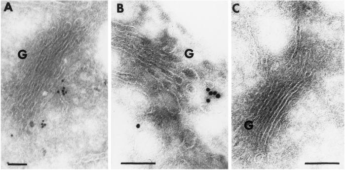
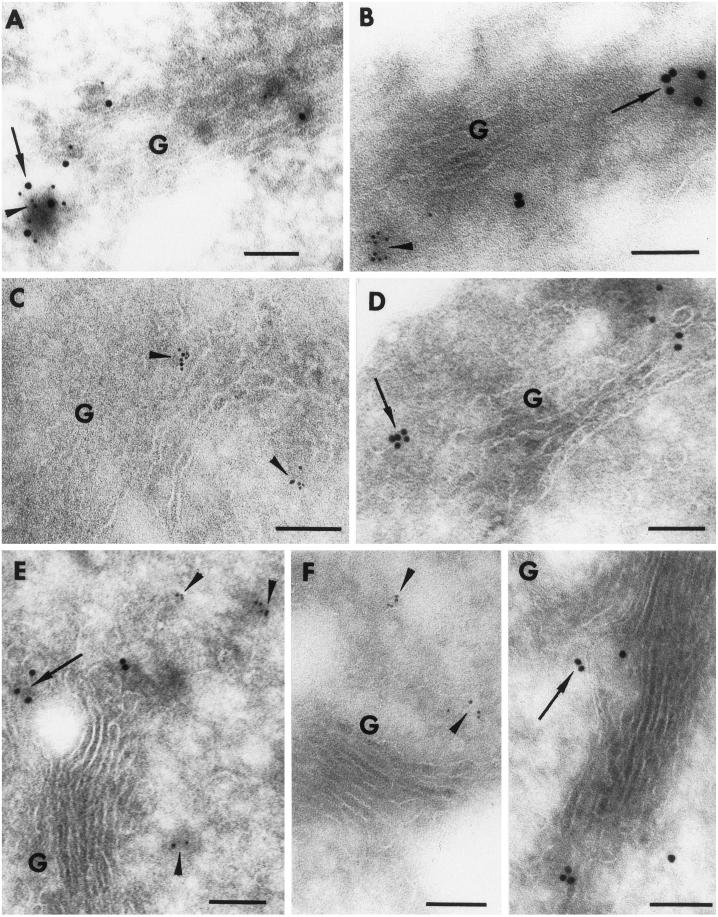
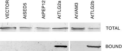
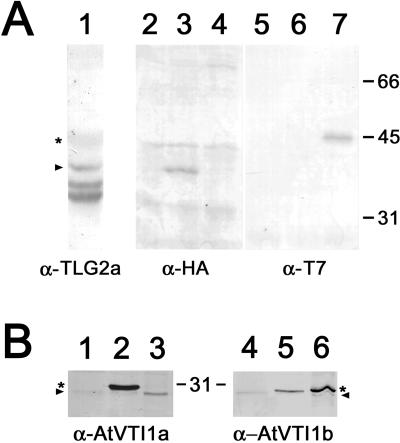
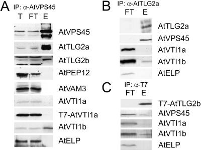
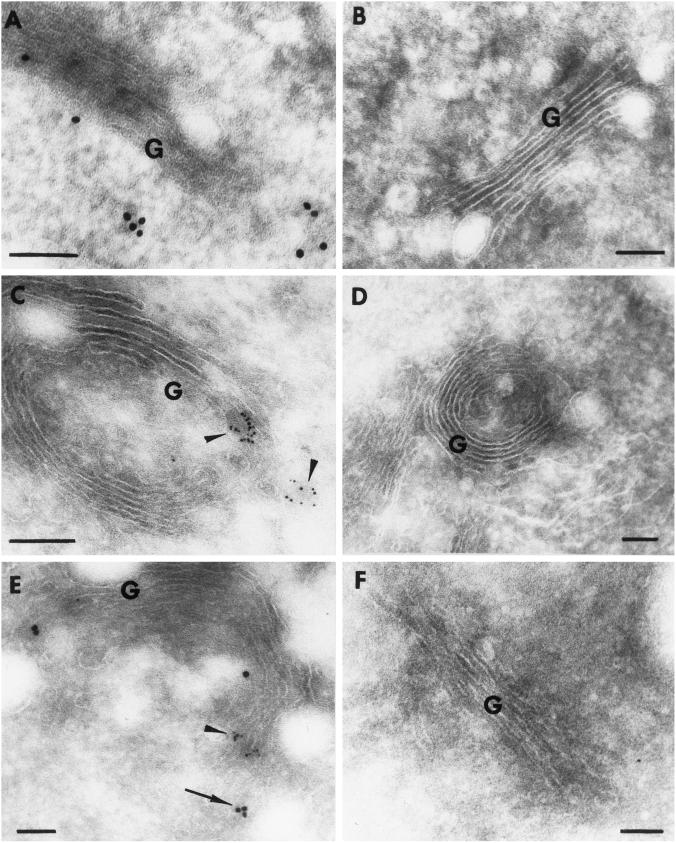
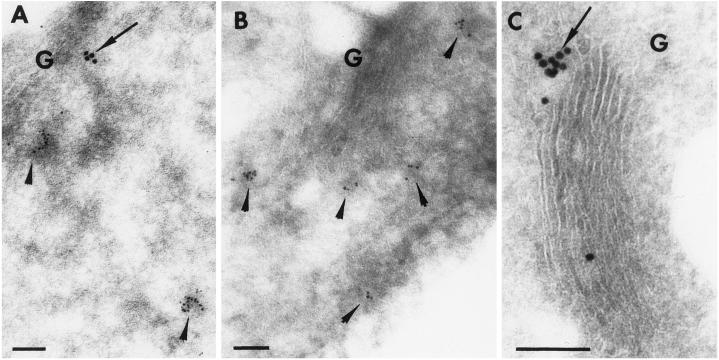
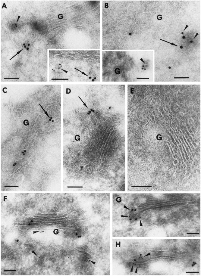
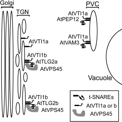
The Sec1p family of proteins are thought to be involved in the regulation of vesicle fusion reactions through interaction with t-SNAREs (target soluble N-ethylmaleimide-sensitive factor attachment protein receptors) at the target membrane. AtVPS45 is a member of this family from Arabidopsis thaliana that we now demonstrate to be present on the trans-Golgi network (TGN), where it colocalizes with the vacuolar cargo receptor AtELP. Unlike yeast Vps45p, AtVPS45 does not interact with, or colocalize with, the prevacuolar t-SNARE AtPEP12. Instead, AtVPS45 interacts with two t-SNAREs, AtTLG2a and AtTLG2b, that show similarity to the yeast t-SNARE Tlg2p. AtTLG2a and -b each colocalize with AtVPS45 at the TGN; however, AtTLG2a is in a different region of the TGN than AtTLG2b by immunogold electron microscopy. Therefore, we propose that complexes containing AtVPS45 and either AtTLG2a or -b define functional subdomains of the TGN and may be required for different trafficking events. Among other Arabidopsis SNAREs, AtVPS45 antibodies preferentially coprecipitate AtVTI1b over the closely related isoform AtVTI1a, implying that AtVTI1a and AtVTI1b also have distinct functions within the cell. These data point to a functional complexity within the plant secretory pathway, where proteins encoded by gene families have specialized functions, rather than functional redundancy.


Figures

Figure 1
Figure 1
Immunolocalization of AtVPS45 on ultrathin cryosections from Arabidopsis roots. (A and B) Sections were incubated with AtVPS45 antibodies followed by biotinylated goat anti-rabbit secondary antibodies and detected with the use of streptavidin conjugated to 10-nm gold particles. (C) Control section probed as for A and B except for the use of preimmune serum in place of the AtVPS45 antibodies. G, Golgi. Bar, 0.1 μm.
Figure 2
Figure 2
AtVPS45 colocalizes with AtELP, and not with AtPEP12, on ultrathin cryosections of Arabidopsis roots. (A and B) Double labeling for AtELP and AtVPS45. Sections were incubated with AtELP antibodies followed by biotinylated goat anti-rabbit secondary antibodies and detected with the use of streptavidin conjugated to 5-nm gold particles. After a second fixation step, the same sections were incubated with antibodies against AtVPS45, then biotinylated goat anti-rabbit antibodies, and detected with the use of streptavidin conjugated to 10-nm gold particles. (C) As for A and B, except preimmune serum was substituted for the AtVPS45 antibodies. (D) As for A and B, except preimmune serum was substituted for AtELP. (E) Double labeling for AtPEP12 and AtVPS45. Sections were labeled according to the protocol described in A and B, with the use of AtPEP12 antibodies followed by 5-nm gold particles and AtVPS45 antibodies followed by 10-nm gold particles. (F) As for E, except preimmune serum was substituted for AtVPS45 antibodies. (G) As for E, except preimmune serum was substituted for AtPEP12 antibodies. G, Golgi. Arrows, 10-nm gold particles (labeling AtVPS45); arrowheads, 5-nm gold particles (labeling AtELP or AtPEP12). Bar, 0.1 μm.
Figure 3
Figure 3
AtVPS45 interacts with AtTLG2a and AtTLG2b in a yeast expression system. Yeast were cotransformed with cDNAs encoding the His-tagged t-SNAREs AtSED5, AtPEP12, AtTLG2a, AtVAM3, or AtTLG2b, or with vector alone as a control, and untagged AtVPS45. The t-SNAREs were isolated from the yeast extracts with the use of a Ni2+-agarose column, and aliquots of the total extracts (TOTAL) and purified t-SNARE complexes (BOUND) were analyzed by SDS-PAGE and immunoblotting with antibodies against AtVPS45.
Figure 4
Figure 4
Characterization of antibodies and epitope tags for AtTLG2a/b and AtVTI1a/b. (A) A microsomal extract of wild-type Arabidopsis (lane 1) was probed with affinity-purified antiserum to AtTLG2a. Full-length AtTLG2a is indicated by the arrowhead, and the smaller bands represent proteolytic breakdown products (see text). Antiserum to AtTLG2a cross-reacts weakly with AtTLG2b, which is indicated by the asterisk. Total protein extracts from either wild type (lanes 2 and 5) or plants expressing either HA-AtTLG2a (lanes 3 and 6) or T7-AtTLG2b (lanes 4 and 7) were probed with antisera specific to the epitope tags. Lanes 2–4 were probed with rabbit anti-HA to indicate HA-AtTLG2a, whereas lanes 5–7 were probed with mouse T7 mAbs to indicate T7-AtTLG2b. (B) Total extracts of wild-type Arabidopsis (lanes 1 and 4), plants expressing T7-AtVTI1a (lanes 2 and 5), or plants expressing HA-AtVTI1b (lanes 3 and 6) were probed with rabbit polyclonal antisera raised to either AtVTI1a (lanes 1–3) or AtVTI1b (lanes 4–6). In each case, the endogenous AtVTI1a or AtVTI1b is indicated by the arrowhead, and the epitope-tagged protein is indicated by the asterisk.
Figure 5
Figure 5
AtTLG2a, AtTLG2b, and AtVTI1b coimmunoprecipitate with the use of AtVPS45 antibodies. (A) Detergent-solubilized membrane preparations from Arabidopsis roots grown in liquid culture were subjected to immunoisolation with the use of AtVPS45 antibodies. Aliquots of the total extract (T), the flow through after immunoprecipitation (FT), and the eluate from the antibody column (E) were analyzed by SDS-PAGE and immunoblotting with the indicated antibodies. In the case of T7-AtTLG2b and T7-AtVTI1a, the immunoprecipitations were performed with the use of tissue from transgenic plants expressing the indicated epitope-tagged protein and probed with the use of mAbs against the T7 tag. (B and C) Detergent-solubilized membranes from wild-type (B) or T7-AtTLG2b–expressing (C) Arabidopsis roots grown in liquid culture were subjected to immunoisolation with AtTLG2a antibodies (B) or T7 mAbs (C). The entire flow through was precipitated by trichloroacetic acid, and equal amounts of flow through and eluate were analyzed by SDS-PAGE and immunoblotting with the indicated antibodies.
Figure 6
Figure 6
AtTLG2a and AtTLG2b localize to different subdomains of the TGN. (A) Ultrathin cryosections of Arabidopsis roots were labeled with AtTLG2a antibodies followed by biotinylated goat anti-rabbit secondary antibodies and detected with the use of streptavidin conjugated to 10-nm gold particles. (B) Control section probed as for A except for the use of preimmune serum in place of the AtTLG2a antibodies. (C) Sections from transgenic plants expressing T7-AtTLG2b were incubated with T7 mAbs followed by a rabbit anti-mouse bridge, biotinylated goat anti-rabbit antibodies, and streptavidin conjugated to 5-nm gold particles. (D) Control section as for C except incubated with 2% BSA in place of the T7 mAbs. (E) Sections from transgenic Arabidopsis producing HA-AtTLG2a and T7-AtTLG2b were incubated with HA polyclonal antibodies followed by biotinylated goat anti-rabbit secondary antibodies and detected with the use of streptavidin conjugated to 10-nm gold particles. After a second fixation step, the same sections were incubated with T7 mAbs, followed by a rabbit anti-mouse bridge and then biotinylated goat anti-rabbit antibodies, and detected with the use of streptavidin conjugated to 5-nm gold particles. (F) Double control probed as for E except for the use of nonimmune serum in place of HA antibodies and 2% BSA in place of T7 antibodies. G, Golgi. Arrow, 10-nm gold particles (labeling HA-AtTLG2a); arrowheads, 5-nm gold particles (labeling T7-AtTLG2b). Bar, 0.1 μm.
Figure 7
Figure 7
AtTLG2a is not present on the PVC. (A) Double labeling was performed on cryosections from Arabidopsis roots as described for Figure 2 with the use of antibodies against AtPEP12 (5 nm gold) and AtTLG2a (10 nm gold). (B) Control labeled as for A except for the use of AtTLG2a preimmune serum in place of anti-AtTLG2a. (C) Control labeled as for A except for the use of AtPEP12 preimmune serum in place of anti-AtPEP12. G, Golgi. Bar, 0.1 μm.
Figure 8
Figure 8
AtTLG2a and AtTLG2b colocalize with AtVPS45. Double labeling was performed as described in Figure 2. (A and inset) AtTLG2a and AtVPS45 on separate parts of the TGN. Cryosections were labeled with AtTLG2a antibodies (5 nm gold) followed by AtVPS45 antibodies (10 nm gold). (B) AtTLG2a and AtVPS45 sometimes colocalize at the TGN. Sections were labeled with AtTLG2a antibodies (10 nm gold) followed by AtVPS45 antibodies (15 nm gold). (B inset) Sections were labeled with AtTLG2a antibodies (5 nm gold) followed by AtVPS45 antibodies (10 nm gold). (C) As for B except AtVPS45 preimmune serum was used in place of anti-AtVPS45. (D) As for A except AtTLG2a preimmune serum was used in place of anti-AtTLG2a. (E) Double control as for A but with both preimmune sera in place of the equivalent antibody. (F–H) Sections from transgenic Arabidopsis expressing T7-tagged AtTLG2b. Labeling was performed as described in Figure 6. (F) T7-AtTLG2b and AtVPS45 on separate parts of the TGN. Sections were labeled with T7 antibodies (5 nm gold) followed by AtVPS45 antibodies (10 nm gold). (G and H) T7-AtTLG2b and AtVPS45 sometimes colocalize at the TGN. Sections were labeled with T7 antibodies (5 nm gold) followed by AtVPS45 antibodies (10 nm gold). G, Golgi. Arrows, 10-nm gold particles (15 nm in B); arrowheads, 5-nm gold particles (10 nm in B). Bar, 0.1 μm.
Figure 9
Figure 9
Summary of results. The t-SNAREs AtTLG2a and AtTLG2b reside on distinct domains of the TGN, where each is found in separate complexes with the Sec1p family member AtVPS45. Unlike the homologous yeast Sec1p family member Vps45p, AtVPS45 is not found on the PVC, nor does it interact with the PVC t-SNAREs AtPEP12 and AtVAM3. Although the highly homologous SNAREs AtVTI1a and AtVTI1b are each found on the TGN, only AtVTI1b is found in AtTLG2a-AtVPS45 and AtTLG2b-AtVPS45 complexes. AtVTI1a has been shown previously to be localized also to the PVC and to be found in complexes with the PVC t-SNAREs AtPEP12 and AtVAM3 (Sanderfoot et al., 1999).

References

    1. Aalto MK, Ronne H, Keränen S. Yeast syntaxins Sso1p and Sso2p belong to a family of related membrane proteins that function in vesicular transport. EMBO J. 1993;12:4095–4104. - PMC - PubMed
    1. Abeliovich H, Darsow T, Emr SD. Cytoplasm to vacuole trafficking of aminopeptidase I requires a t-SNARE-Sec1p complex composed of Tlg2p and Vps45p. EMBO J. 1999;18:6005–6016. - PMC - PubMed
    1. Abeliovich H, Grote E, Novick P, Ferro-Novick S. Tlg2p, a yeast syntaxin homolog that resides on the Golgi and endocytic structures. J Biol Chem. 1998;273:11719–11727. - PubMed
    1. Ahmed SU, Bar-Peled M, Raikhel NV. Cloning and subcellular location of an Arabidopsis receptor-like protein that shares common features with protein-sorting receptors of eukaryotic cells. Plant Physiol. 1997;114:325–336. - PMC - PubMed
    1. Banta LM, Vida TA, Herman PK, Emr SD. Characterization of yeast Vps33p, a protein required for vacuolar protein sorting and vacuole biogenesis. Mol Cell Biol. 1990;10:4638–4649. - PMC - PubMed

Publication types