. 2000 Jul;11(7):2373-85.
doi: 10.1091/mbc.11.7.2373.

The Kar3p kinesin-related protein forms a novel heterodimeric structure with its associated protein Cik1p

Affiliations
Free PMC article

The Kar3p kinesin-related protein forms a novel heterodimeric structure with its associated protein Cik1p

J G Barrett et al. Mol Biol Cell. 2000 Jul.
Free PMC article

Abstract

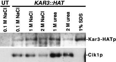
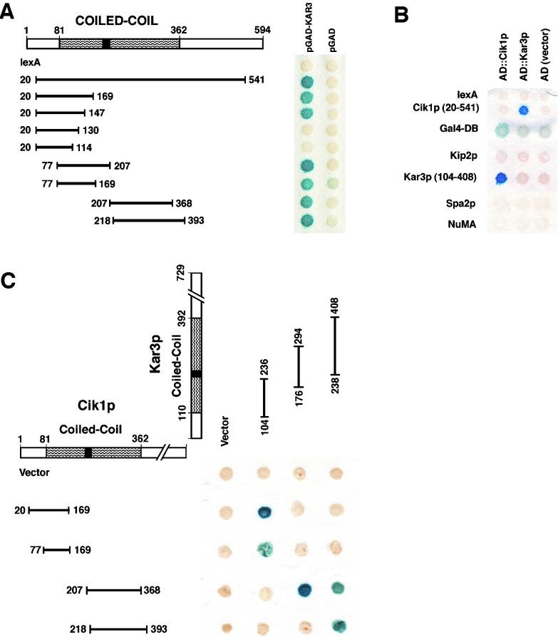
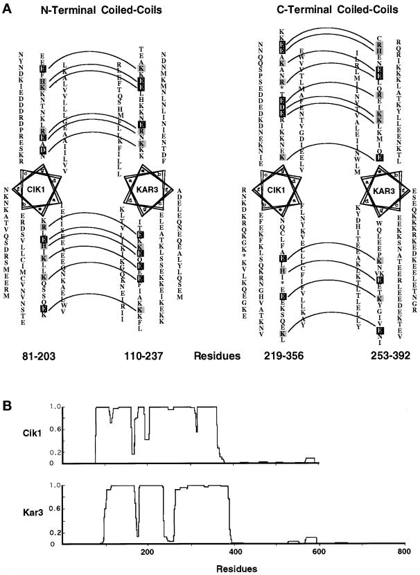
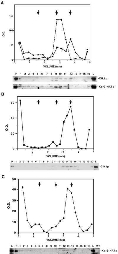
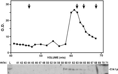
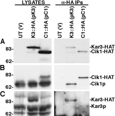
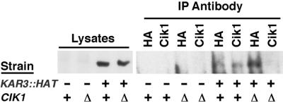
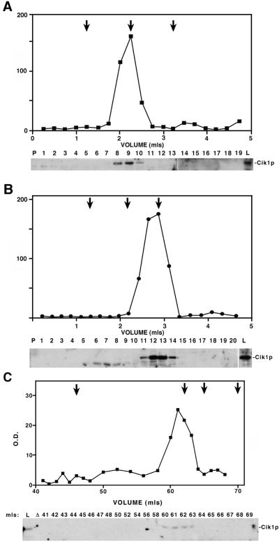
Proteins that physically associate with members of the kinesin superfamily are critical for the functional diversity observed for these microtubule motor proteins. However, quaternary structures of complexes between kinesins and kinesin-associated proteins are poorly defined. We have analyzed the nature of the interaction between the Kar3 motor protein, a minus-end-directed kinesin from yeast, and its associated protein Cik1. Extraction experiments demonstrate that Kar3p and Cik1p are tightly associated. Mapping of the interaction domains of the two proteins by two-hybrid analyses indicates that Kar3p and Cik1p associate in a highly specific manner along the lengths of their respective coiled-coil domains. Sucrose gradient velocity centrifugation and gel filtration experiments were used to determine the size of the Kar3-Cik1 complex from both mating pheromone-treated cells and vegetatively growing cells. These experiments predict a size for this complex that is consistent with that of a heterodimer containing one Kar3p subunit and one Cik1p subunit. Finally, immunoprecipitation of epitope-tagged and untagged proteins confirms that only one subunit of Kar3p and Cik1p are present in the Kar3-Cik1 complex. These findings demonstrate that the Kar3-Cik1 complex has a novel heterodimeric structure not observed previously for kinesin complexes.


Figures

Figure 1
Figure 1
Kar3p and Cik1p are tightly associated. Lysates from an untagged strain (Y1861; UT) or a KAR3::HAT strain (Y1870) were treated with final concentrations of 0.1, 1, or 2 M NaCl, 1 or 2 M urea, or 1% SDS. Lysates were then diluted and immunoprecipitated with anti-HA antibodies. Immunoblot analysis with anti-HA antibodies (top blot) and anti-Cik1p antibodies (bottom blot) were used to detect coimmunoprecipitation of Kar3-HATp and Cik1p from these treated lysates.
Figure 2
Figure 2
Cik1p and Kar3p associate through specific interactions between their coiled-coil domains. (A) Cik1p interacts with Kar3p through its coiled-coil domain. A scheme of the different Cik1p fusions with the lexA DNA-binding domain is shown, with results of the corresponding two-hybrid experiments shown to the right. Amino acids 81–362 represent the long central coiled coil of Cik1p (predicted coiled-coil domains are indicated by curved lines, with breaks indicated by black boxes). Each construct was tested with both the KAR3::AD fusion and with plasmid alone in Y1849. (B) Specificity of the Cik1p-Kar3p interactions. A Cik1p fusion (amino acids 20–541), which interacts with Kar3p, was tested for interaction with full-length Cik1-AD. A Kar3-DBD fusion (amino acids 104–408), which interacts with Cik1p, was tested with full-length Kar3-AD. Cik1-AD and Kar3-AD fusions were also tested for interactions with DBD fusions of the control proteins Kip2p, Spa2p, and NuMA. (C) Kar3p interacts with Cik1p through its coiled-coil domain in a parallel manner. Four of the Cik1-DBD fusions were tested with three Kar3p fusions containing the first half of the Kar3p coiled-coil domain (amino acids 104–236), the middle of the coil (amino acids 176–294) spanning the predicted break (amino acids 237–253), or the second half of the Kar3p coiled-coil domain (amino acids 238–408). In each panel, blue indicates protein-protein interactions resulting in the expression and activity of β-galactosidase, as detected on 5-bromo-4-chloro-3-indolyl-β-d-galactoside (X-Gal)–containing agar plates.
Figure 3
Figure 3
Helical wheel alignments of the coiled-coil domains of Cik1p and Kar3p. (A) Sequence analysis shows how interactions between residues of opposite charges at the e and g positions in the predicted amino-terminal coiled coils of Cik1p and Kar3p (left) and the predicted carboxy-terminal coiled coils of Cik1p and Kar3p (right) might direct the specific interaction between the proteins' coils. Helical wheels were constructed to represent the heptad periodicity of the predicted coils and are arranged so that the hydrophobic a and d residues from each of the coils are across from each other. This allows comparison between the e and g residues of the two proteins that could form electrostatic interactions. When opposite charged residues are present, they are denoted by gray shading (for positively charged) and black shading (for negatively charged), and the pairs are linked by dashed lines. The phase of the heptad repeats was changed at amino acids 293 and 300 for Cik1p and is denoted by asterisks. The phase of the Kar3p repeats was changed once at amino acid 316 and is denoted by having two amino acids in the g position. (B) The predicted coiled coils from Cik1p (top) and Kar3p (bottom) are presented with the use of COILS (version 2.2; Lupas, 1996).
Figure 4
Figure 4
Cik1p and Kar3-HATp cosediment during sucrose density gradient centrifugation with an average S value of 6.26S. Lysates of pheromone-treated cells from wild-type KAR3::HAT (Y1870; A), kar3Δ (Y1700; B), and cik1Δ KAR3::HAT (Y1874; C) strains were separated on 5–20% sucrose gradients. Pellets (P), lysates (L), and proteins from collected fractions (fraction numbers) were analyzed by SDS-PAGE and immunoblots with anti-Cik1p antibodies (A, top blot, and B) and anti-HA antibody (A, bottom blot, and C). Proteins present on immunoblots were quantified with the use of NIH Image software (version 1.59) to determine the relative optical density of the protein of interest in each lane. This value is pictured plotted against the volume eluted: Kar3-HATp (dashed line); Cik1p (solid line). Marker proteins of known S values were run simultaneously with the lysates, and the peaks at which they eluted are represented by arrows: catalase (11.3S; left arrows), aldolase (7.4S; middle arrows), and BSA (4.4S; right arrows).
Figure 5
Figure 5
Gel filtration of the Kar3p-Cik1p complex. Peak sucrose gradient fractions in which Kar3p and Cik1p cofractionated (fractions 9, 10, and 11 from Figure 4A) were pooled and run with protein standards on a Sephacryl S-300 gel filtration column with the use of FPLC. The presence of the Kar3-HATp-Cik1p complex in lysates (L) and collected fractions (fraction numbers) was detected on immunoblots with the use of anti-Cik1p antibodies. Cik1 protein present on immunoblots was quantified with the use of NIH Image software (version 1.59) to determine the relative optical density in each fraction. This value is pictured plotted against the volume eluted. The arrows indicate mobilities of standard proteins of known Rs (from left to right): thyroglobulin (8.5 nm), catalase (5.3 nm), aldolase (4.8 nm), and BSA (3.5 nm).
Figure 6
Figure 6
Kar3p and Cik1p associate with each other but not with themselves in immunoprecipitation experiments. Proteins from lysates of α-factor–treated cells from an untagged strain (Y1861) containing a CEN vector (YCp50) alone [UT (V)], a KAR3::HAT strain (Y1870) containing YCp50 encoding untagged KAR3 [K3::HA(pK3)], and a CIK1::HAT strain (Y2160) containing a YCp50 encoding untagged CIK1 [C1::HA(pC1)] were separated by SDS-PAGE and analyzed by immunoblotting (LYSATES) or first immunoprecipitated with anti-HA antibodies (α-HA IPs). These immunoblots were probed with anti-HA antibodies (A), anti-Cik1p antibodies (B), or anti-Kar3p antibodies (C). The identities of the protein bands in both the lysates and immunoprecipitates are denoted on the right, and the positions of molecular mass markers are shown in kilodaltons on the left.
Figure 7
Figure 7
Kar3-HATp coimmunoprecipitates with Cik1p in vegetatively growing cells. Proteins from lysates of vegetatively growing cells from either untagged strains (wild type [Y1861] and cik1Δ [Y1850]) or KAR3::HAT strains (wild type [Y1870] and cik1Δ [Y1874]) were separated by SDS-PAGE and analyzed by immunoblotting (Lysates) or first immunoprecipitated with anti-HA or anti-Cik1p antibodies (IP Antibody). The genotype of the strain in each lane is denoted under the immunoblot. Immunoblots were probed with anti-HA antibodies.
Figure 8
Figure 8
The Kar3p-Cik1p complex has similar size characteristics in vegetatively growing cells to those exhibited in pheromone-treated cells. (A and B) Sucrose density gradient centrifugation was performed on vegetatively growing wild-type KAR3::HAT (Y1870; A) and kar3Δ (Y1700; B) cell lysates. The Cik1 protein from pellets (P), lysates (L), and collected fractions (fraction numbers) were detected by immunoblots probed with anti-Cik1p antibodies. Cik1p on immunoblots was quantified with the use of NIH Image software (version 1.59) to determine the relative optical density of the protein in each lane. This value is pictured plotted against the volume eluted. Marker proteins of known S values were run simultaneously with the lysates, and the peaks at which they eluted are represented by arrows: catalase (11.3S; left arrows), aldolase (7.4S; middle arrows), and BSA (4.4S; right arrows). (C) Peak Cik1p fractions from the wild-type sucrose gradient experiment (fractions 8, 9, and 10 in A) were pooled and fractionated on a Sephacryl S-300 gel filtrationcolumn with the use of FPLC. Cik1p from collected fractions was analyzed on anti-Cik1p immunoblots, and the measured relative optical density of the protein in each fraction is shown plotted against the volume eluted. The arrows indicate mobilities of standard proteins of known Rs (from left to right): thyroglobulin (8.5 nm), catalase (5.3 nm), aldolase (4.8 nm), and BSA (3.5 nm).
Figure 9
Figure 9
Model of the structure and function of the heterodimeric Kar3p-Cik1p complex in cross-linking and sliding antiparallel microtubules. Kar3p (gray shading) and Cik1p (black shading) interact through an extended coiled-coil stalk domain containing a short break or hinge region. The amino-terminal globular domains of the two proteins (small ovals) act together to bind a microtubule, whereas the motor domain of Kar3p (large ovals with arrows) binds and moves along an antiparallel microtubule toward its minus end. Several Kar3p-Cik1p complexes acting together could pull the minus ends of microtubules toward one another. This activity could drive the nuclear congression step of karyogamy and create the force needed for proper mitotic spindle assembly and/or stability, because both proteins are required for these processes.

References

    1. Arndt K, Pelletier J, Muller K, Alber T, Michnick S, Pluckthun A. A heterodimeric coiled-coil peptide pair selected in vivo from a designed library-versus-library ensemble. J Mol Biol. 2000;295:627–639. - PubMed
    1. Berliner E, Young E, Anderson K, Mahtani H, Gelles J. Failure of single-headed kinesin to track parallel to microtubule protofilaments. Nature. 1995;373:718–721. - PubMed
    1. Bloom GS, Wagner MC, Pfister KK, Brady ST. Native structure and physical properties of bovine brain kinesin and identification of the ATP-binding subunit polypeptide. Biochemistry. 1988;27:3409–3416. - PubMed
    1. Brady S. A novel brain ATPase with properties expected for the fast axonal transport motor. Nature. 1985;317:73–75. - PubMed
    1. Brent R, Ptashne M. A eukaryotic transcriptional activator bearing the DNA specificity of a prokaryotic repressor. Cell. 1985;43:729–736. - PubMed

Publication types

MeSH terms

LinkOut - more resources