. 2000 Jul;123(3):883-94.
doi: 10.1104/pp.123.3.883.

Complementary expression of two plastid-localized sigma-like factors in maize

Affiliations

Complementary expression of two plastid-localized sigma-like factors in maize

S D Lahiri et al. Plant Physiol. 2000 Jul.

Abstract

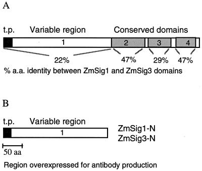
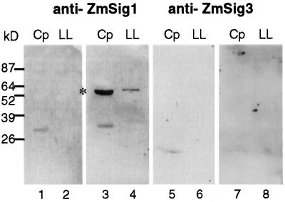
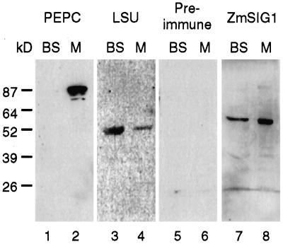
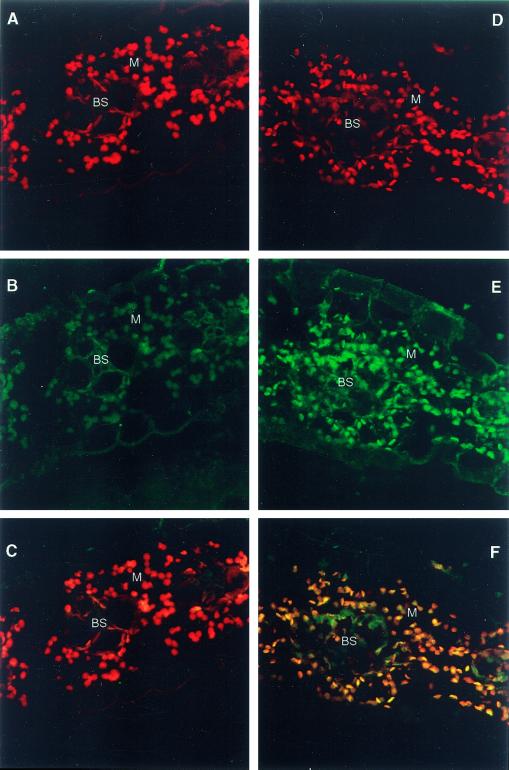
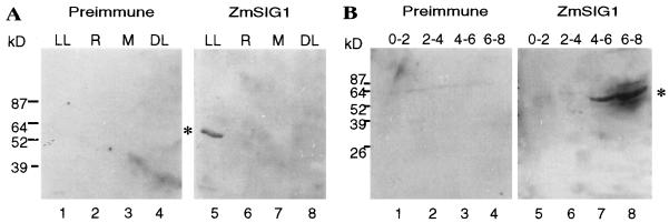
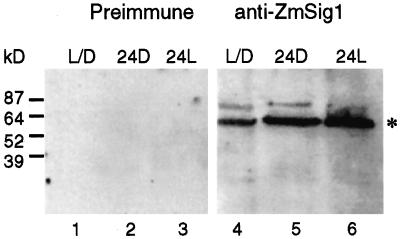
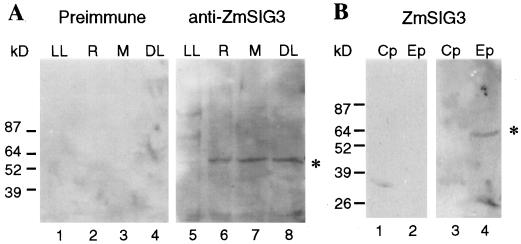
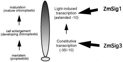
The eubacterial-like RNA polymerase of plastids is composed of organelle-encoded core subunits and nuclear-encoded sigma-factors. Families of sigma-like factors (SLFs) have been identified in several plants, including maize (Zea mays) and Arabidopsis. In vitro import assays determined that at least two of the maize sigma-like proteins have functional chloroplast transit peptides and thus are likely candidates for chloroplast transcriptional regulators. However, the roles of individual SLFs in chloroplast transcription remain to be determined. We have raised antibodies against the unique amino-terminal domains of two maize SLFs, ZmSig1 and ZmSig3, and have used these specific probes to examine the accumulation of each protein in different maize tissues and during chloroplast development. The expression of ZmSig1 is tissue specific and parallels the light-activated chloroplast development program in maize seedling leaves. Its accumulation in mature chloroplasts however, is not affected by subsequent changes in the light regime. It is interesting that the expression profile of ZmSig3 is complementary to that of ZmSig1. It accumulates in non-green tissues, including roots, etiolated seedling leaves, and the basal region of greening seedling leaves. The nonoverlapping expression patterns of these two plastid-localized SLFs suggest that they may direct differential expression of plastid genes during chloroplast development.


Figures

Figure 1
Figure 1
Diagram depicting features of plant polypeptides belonging to the ς-70 family. A, The domains of plant SLFs are indicated: The NH2-terminal transit peptide sequence (t.p.) is indicated by a black box; the variable region 1 is depicted as a white box; conserved regions 2, 3, and 4 are diagrammed as gray boxes. Regions shown are as originally defined in Escherichia coli ς-70 by Lonetto et al. (1992). The percent amino acid identities between the analogous regions in ZmSig1 and ZmSig3 are shown. B, Diagram indicating the regions of ZmSig1 and ZmSig3 that were overexpressed for antibody production. Note that the highly conserved C-terminal region of each protein was not included in the recombinant protein antigens ZmSig1-N and ZmSig3-N.
Figure 2
Figure 2
ZmSig1 is localized to maize leaf chloroplasts. Soluble proteins (20 μg) extracted from intact maize leaf chloroplasts (Cp) and from whole leaf tissue (40 μg, LL) were immunoblotted with either anti-ZmSig1 antisera (lanes 1 and 2, preimmune serum; lanes 3 and 4, immune serum) or with anti-ZmSig3 antisera (lanes 5 and 6, preimmune serum; lanes 7 and 8, immune serum). A chloroplast-localized immunoreactive protein was detected by anti-ZmSig1 as indicated by an asterisk. No immunoreactive proteins were detected in these tissues by anti-ZmSig3 antibodies. The positions of molecular mass standards are indicated to the left.
Figure 3
Figure 3
ZmSig1 is expressed in both bundle sheath and mesophyll cells of the maize leaf. In each panel 40 μg of proteins extracted from purified bundle sheath (BS) and mesophyll (M) cells was separated on SDS-PAGE and immunoblotted with the following antibodies: lanes 1 and 2, antibodies against maize PEPC; lanes 3 and 4, antibodies against maize Rubisco LSU; lanes 5 and 6, anti-ZmSig1 preimmune serum; and lanes 7 and 8, anti-ZmSig1 immune serum. The immunoreactive ZmSig1 protein was detected in pure preparations of bundle sheath cells (lane 7) as well as in the mesophyll cell preparation (lane 8). Note that, based on the LSU signals, the mesophyll cells were slightly contaminated with bundle sheath cell proteins (lane 3 versus lane 4). The positions of molecular mass standards are indicated to the left.
Figure 4
Figure 4
Immunocalization of ZmSig1 in mesophyll (M) and bundle-sheath (BS) cells of a maize leaf. Maize leaf sections from the mid-leaf region of 21-d-old seedlings were probed with preimmune (A–C) or anti-ZmSig1 (D–F) antisera and subsequently detected with an anti-IgG secondary antibody conjugated to a fluorescent probe. The signals were visualized using a Bio-Rad MRC600 confocal microscope. The red signals in A and D are due to chlorophyll autofluorescence within the chloroplast (excitation at 647 nm and emission at 666 nm); whereas the green fluorescence in B and E (excitation at 488 nm and emission at 520 nm) is due to the secondary antibody and thus represents immunoreactive protein. The immunoreactive protein is localized mainly in chloroplasts, since merging the red and green signals results in a yellow image (F). Note that, since bundle sheath chloroplasts have a less intense autofluorescent (red) signal than mesophyll cell chloroplasts, the immunoreactive protein signal in BS cells appears green in the merged image (F).
Figure 5
Figure 5
Tissue-specific and developmentally regulated expression of the maize ZmSig1 protein. A, Total proteins were extracted from the top portions (2–7 cm above the leaf base) of 4-d-old greening maize seedlings (LL), from the roots of the same seedlings (R), from the leaf base (0–2 cm region) of the seedling leaves (M), and from the etiolated leaves of 4-d-old seedlings grown in complete darkness (DL). Forty micrograms of protein was separated and immunoblotted with either preimmune (lanes 1–4) or immune (lanes 5–8) antisera against ZmSig1. The immunoreactive 60-kD ZmSig1 protein was detected only in the upper regions of light-grown leaves (asterisk). B, Leaves from 5-d-old light-grown maize seedlings were cut into sections that measured 0 to 2, 2 to 4, 4 to 6, or 6 to 8 cm from the leaf base. Total proteins (40 μg) extracted from each section were immunoblotted with either preimmune (lanes 1–4) or immune (lanes 5–8) antisera against ZmSig1. The immunoreactive 60-kD ZmSig1 protein was detected only in the upper regions of light-grown leaves, from 4 to 8 cm above the leaf base (asterisk). This discrete expression profile indicates that ZmSig1 accumulates preferentially in mature chloroplasts. The positions of molecular mass standards are indicated.
Figure 6
Figure 6
The accumulation of ZmSig1 protein in mature chloroplasts is not influenced by light. Proteins were extracted from the upper region of leaf tissues of seedlings grown for 7 d in cycling light (L/D), from a similar batch of seedlings transferred to complete darkness for 24 h (24D), and from the same batch of seedlings transferred from the 24-h dark treatment to constant illumination for 24 h (24L). The tissues were harvested at the same time of day (10:30 am) to avoid possible circadian fluctuations in protein accumulation. Extracted proteins (40 μg) were immunoblotted with either preimmune sera (lanes 1–3) or anti-ZmSig1 antisera (lanes 4–6). No dramatic changes in levels of ZmSig1 protein (asterisk) were detected. The positions of molecular mass standards are indicated to the left.
Figure 7
Figure 7
ZmSig3 accumulates in non-green plastids. A, Total proteins (40 μg) from leaf sections 2 to 7 cm above the base of 4-d-old greening maize seedlings (LL), from the roots of the same seedlings (R), from the leaf base of the seedling leaves (M), and from leaves of 4-d-old etiolated seedlings (DL) were immunoblotted with either preimmune (lanes 1–4) or immune (lanes 5–8) antisera against ZmSig3. The immunoreactive 62-kD ZmSig3 protein was detected in the non-green tissues (asterisk) but not in the upper portion of the light-grown leaves. B, Total proteins (25 μg) extracted from chloroplasts (Cp) purified from light-grown leaves, or etioplasts (Ep) purified from etiolated seedling leaves were immunoblotted with either preimmune sera (lanes 1 and 2) or immune sera raised against ZmSig3 (lanes 3 and 4). The immunoblot shows that ZmSig3 protein accumulates in etioplasts (asterisk) rather than chloroplasts. The positions of molecular mass standards are indicated to the left.
Figure 8
Figure 8
A model for SLF function in the developing maize leaf. A maize seedling leaf is diagrammed with three different zones of development indicated (based on Mullet, 1993): the meristematic region at the leaf base, containing proplastids; the zone of cell enlargement, containing early developing chloroplasts; and the maturation zone in the upper region of the leaf, containing mature chloroplasts. The PEP RNA polymerase promoter usage and light dependence derived from experiments with wheat seedlings (Satoh et al., 1999) are indicated to the right. We speculate that ZmSig1, due to its conservation of region 2.5, should be capable of recognizing extended −10 promoters and thus may confer this property upon the PEP RNA polymerase located in the zone of mature chloroplasts. In contrast ZmSig3, whose expression is restricted to the meristematic zone, does not have region 2.5 and thus may restrict the PEP RNA polymerase in developing chloroplasts to recognize promoters containing a −35 sequence element.

Similar articles

Cited by

References

    1. Allison LA (2000) The role of ς factors in plastid transcription. Biochimie (in press) - PubMed
    1. Barne KA, Bown JA, Busby SJW, Minchin SD. Region 2.5 of the Escherichia coli RNA polymerase ς70subunit is responsible for the recognition of the 'extended −10′ motif at promoters. EMBO J. 1997;16:4030–4040. - PMC - PubMed
    1. Baumgartner BJ, Rapp JC, Mullet JE. Plastid transcription activity and DNA copy number increase early in barley chloroplast development. Plant Physiol. 1989;89:1011–1018. - PMC - PubMed
    1. Baumgartner BJ, Rapp JC, Mullet JE. Plastid genes encoding the transcription/translation apparatus are differentially transcribed early in barley (Hordeum vulgare) chloroplast development. Plant Physiol. 1993;101:781–791. - PMC - PubMed
    1. Bensen RJ, Johal GS, Crane VC, Tossberg JT, Schnable PS, Meeley RB, Briggs SP. Cloning and characterization of the maize An1gene. Plant Cell. 1995;7:75–84. - PMC - PubMed

Publication types

LinkOut - more resources