. 2000 Jul;123(3):905-15.
doi: 10.1104/pp.123.3.905.

CHRK1, a chitinase-related receptor-like kinase in tobacco

Affiliations

CHRK1, a chitinase-related receptor-like kinase in tobacco

Y S Kim et al. Plant Physiol. 2000 Jul.

Abstract

A cDNA encoding a chitinase-related receptor-like kinase, designated CHRK1, was isolated from tobacco (Nicotiana tabacum). The C-terminal kinase domain (KD) of CHRK1 contained all of the conserved amino acids of serine/threonine protein kinases. The putative extracellular domain was closely related to the class V chitinase of tobacco and to microbial chitinases. CHRK1 mRNA accumulation was strongly stimulated by infection with fungal pathogen and tobacco mosaic virus. Amino acid-sequence analysis revealed that the chitinase-like domain of CHRK1 lacked the essential glutamic acid residue required for chitinase activity. The recombinant chitinase-like domain did not show any catalytic activity for either oligomeric or polymeric chitin substrates. The recombinant KD of CHRK1 exhibited autophosphorylation, but the mutant KD with a mutation in the essential ATP-binding site did not, suggesting that CHRK1 encoded a functional kinase. CHRK1 was detected in membrane fractions of tobacco BY2 cells. Furthermore, CHRK1-GFP fusion protein was localized in plasma membranes when it was expressed in animal cells. This is the first report of a new type of receptor-like kinase containing a chitinase-like sequence in the putative extracellular domain.


Figures
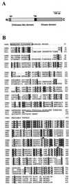
Figure 1
Figure 1
Structure and amino acid sequence comparison of CHRK1. A, Schematic representation of the CHRK1 protein. The signal peptide (SP), the transmembrane region (TM), the putative extracellular domain, and the KD are indicated. B, Deduced amino acid sequence of CHRK1 and alignment with related sequences. The deduced amino acid sequence of the CD of CHRK1 was aligned with the sequences from class V chitinase (CHIV) from tobacco (Melchers et al., 1994) and chitinase A1 (CHIA) from B. circulans (Watanabe et al., 1990). The predicted KD was aligned with the KD of the plant RLKs, SRK6 of Brassica (Stein et al., 1991), and ARK3 (Dwyer et al., 1994) and PR5K (Wang et al., 1996) of Arabidopsis. The number on the right indicates the amino acid residues. Gaps, which were introduced to maximize alignment, are indicated by dots. Residues conserved among all sequences compared here are highlighted in reverse contrast letters. Asterisks indicate the three conserved residues that are important for chitinase activity. Crosses (+) indicate residues that are conserved in all Ser/Thr-type kinases. Asterisks and crosses were written below the relevant amino acids. The putative signal peptide is boxed. The transmembrane region is indicated with double underlines. The amino acid residues corresponding to the degenerate oligonucleotides for PCR amplification are marked with overlines and arrows.
Figure 2
Figure 2
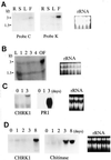
DNA gel-blot analysis of CHRK1 gene. A, Genomic DNA gel- blot analysis. DNA gel blots were hybridized with the chitinase (C) or the kinase (K) probe. The DNA size markers are indicated in kb. Coding regions of CHRK1 cDNA are boxed. E, EcoRI; B, BglII; P, PstI; N, NdeI. B, Genomic Southern-blot analysis of CHRK1-related sequences in other plant species. Each lane represents 10 μg of genomic DNA fragmented with EcoRI. The blot was hybridized with the kinase probe. The DNA size markers are indicated in kb.
Figure 3
Figure 3
Expression of CHRK1 mRNA. A, Tissue-specific expression. Each lane represents 50 μg of total RNA from roots (R), stems (S), leaves (L), or flowers (F). The amount of ethidium bromide-stained rRNA was shown to verify equal loading of RNA in each lane. B, Expression of CHRK1 mRNA during flower development. Fifty micrograms of total RNA of flowers from stage 1 to open flower stage, and from leaves (L) is represented in each lane. The five developmental stages are defined by bud size: <1 cm, stage 1; 1 to 2 cm, stage 2; 2 to 3 cm, stage 3; 3 to 4 cm, stage 4; open flower, stage OF. The K probe was used. C, Expression of CHRK1 mRNA in response to TMV infection. Fifty micrograms of total RNA was used in each lane. Lane 1 contains RNA from uninfected leaves; lane 2 contains RNA from leaves 1 d after infection; lane 3 contains RNA from leaves 3 d after infection. Duplicate membranes were hybridized with PR-1a probe as a control. The size of the PR1 transcripts is approximately 0.9 kb. The K probe was used. D, Expression of CHRK1 mRNA in response to fungal pathogen infection. Young tobacco plants were inoculated through roots with P. parasitica, which causes black shank disease in tobacco. Total RNA was prepared from leaves collected at 0, 1, 2, 3, and 8 d after infection. Fifty micrograms of total RNA was used in each lane. The K probe was used. Duplicate membranes were hybridized with chitinase probe as a control. The size of the chitinase transcripts is approximately 1.2 kb.
Figure 4
Figure 4
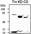
Expression of the CD and the KD of CHRK1 in E. coli. The CD and KD of CHRK1 were expressed in E. coli as fusion proteins with thioredoxin using the pET32a vector. The protein profiles after purification were analyzed by SDS-PAGE. Arrowheads indicate the electrophoretic positions of thioredoxin (Trx), thioredoxin-KD, and thioredoxin-CD proteins. The sizes of molecular mass markers are also indicated.
Figure 5
Figure 5
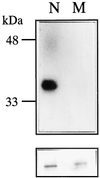
Autophosphorylation of the recombinant KD of CHRK1. Top, Autoradiography; Bottom, Coomassie Blue-stained gel showing the relative amounts of the recombinant proteins. Wild-type (N) and mutant (M) forms of the KD are indicated. In the mutant kinase the essential ATP-binding site, Lys-449, was mutated to Asn.
Figure 6
Figure 6
Sequence comparison of the conserved residues of chitinases with those of the CHRK1 CD. Three residues conserved in plant and microbial chitinases identified by Watanabe et al. (1993) are shown in bold. The sources of other chitinase sequences are basic and acidic class III chitinases from tobacco (Lawyton et al., 1992); class V chitinase from tobacco (Melchers et al., 1994); chitinase A1 from Bacillus subtilis (Watanabe et al., 1990); chitinase from Saccharomyces cerevisiae (Kuranda and Robbins, 1991); HC gp-39 (Hakala et al., 1993); and CHRK1.
Figure 7
Figure 7
Immunodetection of CHRK1 protein. A, Immunoblotting of the recombinant CD with anti-CHRK1 antiserum. Ten micrograms of protein extracts from uninduced (UI) and induced (I) E. coli cells that carry the pET32a-CD plasmid was subjected to immunoblotting. The recombinant CHRK1 is marked with the arrowhead. B, Immunodetection of CHRK1 proteins in membranes of tobacco BY2 cells. Thirty micrograms of soluble (S) and membrane (M) proteins from BY2 cells was subjected to immunoblotting. The CHRK1 protein is marked with the arrowhead. Molecular mass markers are indicated in kD. C, Immunodetection of CHRK1 protein expressed in animal cells. CHRK1 protein was transiently expressed in human 293T cells under the control of CMV promoter. Thirty micrograms of S and M proteins isolated from transfected cells with pCMV or pCMV-CHRK1 was subjected to immunoblotting. The expressed CHRK1 protein is marked with the arrowhead. Molecular mass markers are indicated in kD.
Figure 8
Figure 8
Membrane localization of the CHRK1-GFP fusion protein expressed in animal cells. The CHRK1-GFP and GFP alone were transiently expressed in HeLa cells. The left and the right panel represent light microscopic image and fluorescent microscopic image of individual cells, respectively. A fluorescence filter set 09 (Zeiss, Jena, Germany) was used to observe the fluorescence signal.

Similar articles

Cited by

References

    1. Baureithel K, Felix G, Boller T. Specific, high affinity binding of chitin fragments to tomato cells and membranes: competitive inhibition of binding by derivatives of chitooligosaccharides and a Nod factor of Rhizobium. J Biol Chem. 1994;269:17931–17938. - PubMed
    1. Becraft PW, Stinard PS, McCarty DR. CRINKLY4: a TNFR-like receptor kinase involved in maize epidermal differentiation. Science. 1996;273:1406–1409. - PubMed
    1. Benhamou N. Elicitor-induced plant defense pathways. Trends Plant Sci. 1996;1:233–240.
    1. Bent AF. Plant disease resistance genes: function meets structure. Plant Cell. 1996;8:1757–1771. - PMC - PubMed
    1. Bollag DM, Rozycki MD, Edelstein SJ. Protein Methods. New York: Wiley-Liss; 1996.

Publication types

MeSH terms

LinkOut - more resources