

Identification of phloem involved in assimilate loading in leaves by the activity of the galactinol synthase promoter
- PMID: 10889241
- PMCID: PMC59055
- DOI: 10.1104/pp.123.3.929
Identification of phloem involved in assimilate loading in leaves by the activity of the galactinol synthase promoter
Abstract
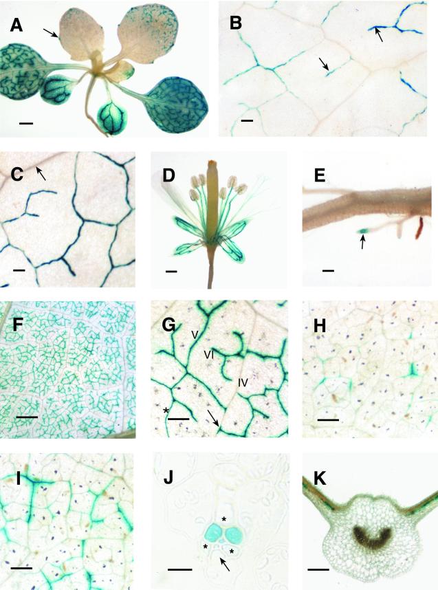
The definition of "minor" veins in leaves is arbitrary and of uncertain biological significance. Generally, the term refers to the smallest vein classes in the leaf, believed to function in phloem loading. We found that a galactinol synthase promoter, cloned from melon (Cucumis melo), directs expression of the gusA gene to the smallest veins of mature Arabidopsis and cultivated tobacco (Nicotiana tabacum) leaves. This expression pattern is consistent with the role of galactinol synthase in sugar synthesis and phloem loading in cucurbits. The expression pattern in tobacco is especially noteworthy since galactinol is not synthesized in the leaves of this plant. Also, we unexpectedly found that expression in tobacco is limited to two of three companion cells in class-V veins, which are the most extensive in the leaf. Thus, the "minor" vein system is defined and regulated at the genetic level, and there is heterogeneity of response to this system by different companion cells of the same vein.
Figures
Similar articles
-
Functional and phylogenetic analyses of a conserved regulatory program in the phloem of minor veins.Plant Physiol. 2003 Nov;133(3):1229-39. doi: 10.1104/pp.103.027714. Epub 2003 Oct 2. Plant Physiol. 2003. PMID: 14526110 Free PMC article.
-
FLOWERING LOCUS T mRNA is synthesized in specialized companion cells in Arabidopsis and Maryland Mammoth tobacco leaf veins.Proc Natl Acad Sci U S A. 2018 Mar 13;115(11):2830-2835. doi: 10.1073/pnas.1719455115. Epub 2018 Feb 26. Proc Natl Acad Sci U S A. 2018. PMID: 29483267 Free PMC article.
-
Functional characterization of the Arabidopsis AtSUC2 Sucrose/H+ symporter by tissue-specific complementation reveals an essential role in phloem loading but not in long-distance transport.Plant Physiol. 2008 Sep;148(1):200-11. doi: 10.1104/pp.108.124776. Epub 2008 Jul 23. Plant Physiol. 2008. PMID: 18650401 Free PMC article.
-
Structural and functional vein maturation in developing tobacco leaves in relation to AtSUC2 promoter activity.Plant Physiol. 2003 Apr;131(4):1555-65. doi: 10.1104/pp.102.016022. Plant Physiol. 2003. PMID: 12692315 Free PMC article.
-
Foliar sieve elements: Nexus of the leaf.J Plant Physiol. 2022 Feb;269:153601. doi: 10.1016/j.jplph.2021.153601. Epub 2021 Dec 17. J Plant Physiol. 2022. PMID: 34953412 Review.
Cited by
-
Modeling the parameters for plasmodesmal sugar filtering in active symplasmic phloem loaders.Front Plant Sci. 2013 Jun 19;4:207. doi: 10.3389/fpls.2013.00207. eCollection 2013. Front Plant Sci. 2013. PMID: 23802006 Free PMC article.
-
Symplastic continuity between companion cells and the translocation stream: long-distance transport is controlled by retention and retrieval mechanisms in the phloem.Plant Physiol. 2003 Apr;131(4):1518-28. doi: 10.1104/pp.012054. Plant Physiol. 2003. PMID: 12692312 Free PMC article.
-
Arabidopsis POLYOL TRANSPORTER5, a new member of the monosaccharide transporter-like superfamily, mediates H+-Symport of numerous substrates, including myo-inositol, glycerol, and ribose.Plant Cell. 2005 Jan;17(1):204-18. doi: 10.1105/tpc.104.026641. Epub 2004 Dec 14. Plant Cell. 2005. PMID: 15598803 Free PMC article.
-
Significance of galactinol and raffinose family oligosaccharide synthesis in plants.Front Plant Sci. 2015 Aug 26;6:656. doi: 10.3389/fpls.2015.00656. eCollection 2015. Front Plant Sci. 2015. PMID: 26379684 Free PMC article. Review.
-
Arabidopsis INOSITOL TRANSPORTER2 mediates H+ symport of different inositol epimers and derivatives across the plasma membrane.Plant Physiol. 2007 Dec;145(4):1395-407. doi: 10.1104/pp.107.109033. Epub 2007 Oct 19. Plant Physiol. 2007. PMID: 17951450 Free PMC article.
References
-
- Ausubel FM, Brent R, Kingston RE, Moore DD, Seidman JG, Smith JA, Struhl K. Current Protocols in Molecular Biology. New York: John Wiley & Sons; 1995.
-
- Beebe DU, Turgeon R. Localization of galactinol, raffinose, and stachyose synthesis in Cucurbita pepo leaves. Planta. 1992;188:354–361. - PubMed
-
- Bernatzky R, Tanksley SD. Genetics of actin-related sequences in tomato. Theor Appl Genet. 1986;72:314–321. - PubMed
-
- Bingham PM, Levis R, Rubin GM. Cloning of DNA sequences from the white locus of D. melanogaster by a novel and general method. Cell. 1981;25:693–704. - PubMed
-
- Caissard JC, Guivarc'h A, Rembur J, Abdelkrim A, Chriqui D. Spurious localizations of diX-indigo microcrystals generated by the histochemical GUS assay. Transgenic Res. 1994;3:176–181.
Publication types
MeSH terms
Substances
LinkOut - more resources
Full Text Sources
