

Recent advances in immunohistochemistry in the diagnosis of ovarian neoplasms
- PMID: 10889812
- PMCID: PMC1731190
- DOI: 10.1136/jcp.53.5.327
Recent advances in immunohistochemistry in the diagnosis of ovarian neoplasms
Abstract
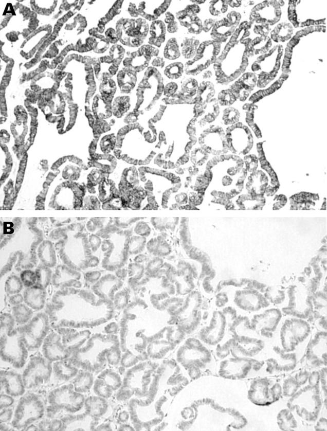
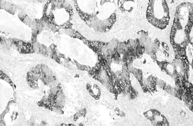
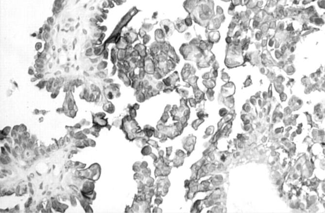
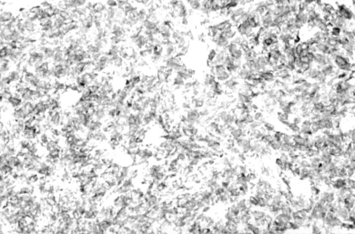
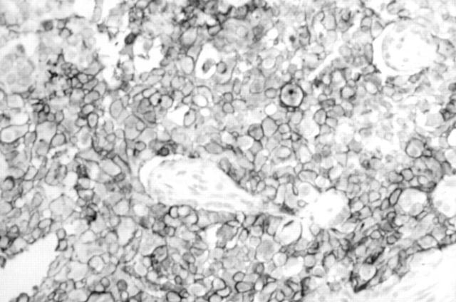
This leader reviews recent advances in immunohistochemistry that are useful in the diagnosis of ovarian neoplasms. These include the value of different anticytokeratin antibodies in the distinction between a primary ovarian adenocarcinoma and a metastatic adenocarcinoma, especially of colorectal origin. These antibodies have also helped to clarify the origin of the peritoneal disease in most cases of pseudomyxoma peritonei. The value of antibodies against so called tumour specific antigens, such as CA125 and HAM56, in determining the ovarian origin of an adenocarcinoma is also reviewed. In recent years, several studies have investigated the value of a variety of monoclonal antibodies in the diagnosis of ovarian sex cord stromal tumours and in the distinction between these neoplasms and their histological mimics. These antibodies include those directed against inhibin, CD99, Mullerian inhibiting substance, relaxin like factor, melan A, and calretinin. Of these, anti-alpha inhibin appears to be of most diagnostic value. It is stressed that these antibodies should always be used as part of a larger panel and not in isolation.
Figures
References
Publication types
MeSH terms
Substances
LinkOut - more resources
Full Text Sources
Other Literature Sources
Medical
Research Materials
Miscellaneous