. 2000 Jul 18;97(15):8352-7.
doi: 10.1073/pnas.140021997.

Phosphate is a specific signal for induction of osteopontin gene expression

Affiliations

Phosphate is a specific signal for induction of osteopontin gene expression

G R Beck Jr et al. Proc Natl Acad Sci U S A. .

Abstract

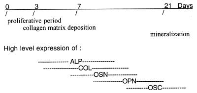
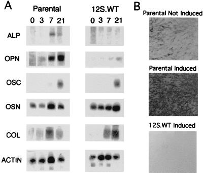
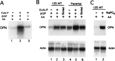
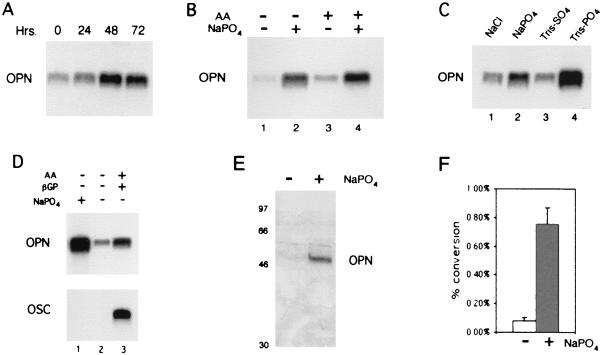
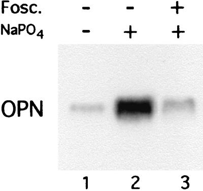
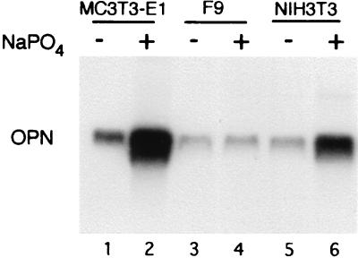
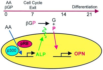
Osteopontin is a phosphorylated glycoprotein secreted to the mineralizing extracellular matrix by osteoblasts during bone development. It is believed to facilitate the attachment of osteoblasts and osteoclasts to the extracellular matrix, allowing them to perform their respective functions during osteogenesis. Several other functions have been suggested for this protein, and its up-regulation is associated with various disease states related to calcification, including arterial plaque formation and the formation of kidney stones. Although expression of this gene has been demonstrated in multiple tissues, its regulation is not well understood. Our previous studies on the roles of the retinoblastoma protein (pRB) and p300/CBP in the regulation of osteoblast differentiation revealed a link between osteopontin induction and the synthesis of alkaline phosphatase. In this paper, we describe results specifically linking induction of osteopontin to the enzymatic activity of alkaline phosphatase in the medium, which results in the generation of free phosphate. This elevation of free phosphate in the medium is sufficient to signal induction of osteopontin RNA and protein. The strong and specific induction of osteopontin in direct response to increased phosphate levels provides a mechanism to explain how expression of this product is normally regulated in bone and suggests how it may become up-regulated in damaged tissue.


Figures

Figure 1
Figure 1
Temporal expression pattern of markers typical of osteoblast development in MC3T3-E1 cells. The cells progress through three general phases: (i) proliferation, (ii) extracellular matrix deposition and maturation, and (iii) mineralization. High-level expression of genes thought to contribute to the differentiated state of osteoblasts occurs at discrete time points during the differentiation process. Alkaline phosphatase (ALP), α1-collagen (COL), and osteonectin (OSN) are expressed at high levels near the end of the proliferative period and during the period of extracellular matrix deposition and maturation. Genes expressed at or near the time of mineralization include osteopontin (OPN) and osteocalcin (OSC).
Figure 2
Figure 2
Characterization of the 12S.WT E1A-expressing cell line. (A) Northern blot characterization of differentiation markers in MC3T3-E1 cell lines. Cells were cultured with differentiation media (see Materials and Methods) and total RNA was prepared at days 0, 3, 7, and 21. The same blot was hybridized sequentially with cDNA probes to alkaline phosphatase (ALP), osteopontin (OPN), osteocalcin (OSC), osteonectin (OSN), α1-collagen (COL), and β-actin (actin), and is described in more detail in ref. . (B) In situ analysis of alkaline phosphatase activity. The cells were cultured in a six-well plate and stained with 5-bromo-4-chloro-3-indolyl phosphate/nitroblue tetrazolium (BCIP/NBT) as described previously (9). Parental-not induced, growth medium; parental-induced, differentiation medium; 12S.WT-induced, differentiation medium. [Reproduced from ref. with permission of John Wiley & Sons, Inc. (copyright 1998 Wiley–Liss, Inc.), http://www.interscience.wiley.com/jpages/0730-23121.]
Figure 3
Figure 3
Northern blot analysis of osteopontin expression after addition of exogenous alkaline phosphatase (ExALP) or sodium phosphate. (A) The 12S.WT.G1 cell line was cultured for 21 days with growth medium supplemented with ExALP, β-glycerol phosphate (βGP), or ascorbic acid (AA) as stated above each lane. (B) The 12S.WT.G1 and parental (MC3T3-E1) cell lines were cultured with growth medium and AA, supplemented with ExALP and βGP or a solution in which ExALP and βGP were incubated and then boiled to inactive the enzyme (Boiled), as stated above each lane. (C) The 12S.WT.G1 cell line was cultured for 21 days in the presence of AA and in the presence or absence of 10 mM sodium phosphate (NaPO4). Blots were hybridized sequentially with cDNA probes to osteopontin (OPN) and β-actin.
Figure 4
Figure 4
Properties of phosphate-signal-regulated osteopontin expression. (A) Northern blot (NB) analysis of 12S.WT.G1 cells grown in the presence of ascorbic acid (AA) and treated with 10 mM sodium phosphate (NaPO4) for 0, 24, 48, and 72 h before harvesting the cells at day 11 for RNA isolation. (B) NB analysis of the parental cells grown in the absence (lanes 1 and 2) or presence (lanes 3 and 4) of AA and then treated (lanes 2 and 4) with 10 mM sodium phosphate for 48 h. The cells were harvested for RNA analysis at day 11. (C) NB analysis of the parental cells given growth medium for 4 days postconfluency and then treated with 10 mM NaCl, 10 mM sodium phosphate, 10 mM Tris-sulfate (TrisSO4), or 10 mM Tris-phosphate (TrisPO4) for 60 h and harvested for RNA analysis. (D) NB analysis of preconfluent MC3T3-E1 (lanes 1 and 2) and day 21 (lane 3) probed with osteopontin (OPN) and osteocalcin (OSC). Treatment conditions are stated above each lane. (E) Western blot analysis of osteopontin in response to 10 mM sodium phosphate. Preconfluent MC3T3-E1 were treated in the presence or absence of 10 mM sodium phosphate for 48 h and analyzed by PAGE. Membranes were probed with the osteopontin-specific antibody LF-123. (F) CAT assay analysis of the osteopontin promoter in response to 10 mM sodium phosphate. Transiently transfected cells were treated with either sodium sulfate (white bar) or sodium phosphate (shaded bar). The graph shows the percentage conversion of chloramphenicol to acetylated forms.
Figure 5
Figure 5
The phosphate transport inhibitor foscarnet blocks up-regulation of osteopontin in response to phosphate. MC3T3-E1 cells 4 days postconfluency were pretreated with 300 μM foscarnet (lane 3) for 3 h and then treated with 10 mM sodium phosphate (NaPO4) for 72 h (lanes 2 and 3) in the continued presence of foscarnet (lane 3) and analyzed by Northern analysis.
Figure 6
Figure 6
Northern blot analysis of osteopontin (OPN) expression in various cell types. Preconfluent MC3T3-E1, NIH 3T3, and F9 cells were treated with 10 mM sodium phosphate (NaPO4) for 48 h and harvested for RNA analysis.
Figure 7
Figure 7
Schematic representation of the coordination of alkaline phosphatase activity and osteopontin levels in osteoblasts. Alkaline phosphatase (ALP) activity is induced by ascorbic acid (AA) in a process that requires the p300/CBP and pRB families. ALP then is localized to the plasma membrane and oriented such that its catalytic subunit is ectoplasmic. Within the extracellular environment, ALP cleaves a phosphate (P) from β-glycerol phosphate (βGP) to leave free glycerol (G). The free phosphate enters the cell through a Na-dependent phosphate transporter, which is subject to inhibition by foscarnet. Transport of the phosphate results in the up-regulation of osteopontin (OPN) RNA levels. During the differentiation process, the requirement for the pRB and p300 families ensures that induction of the early differentiation marker, alkaline phosphatase, does not begin until proliferation has shut down, and expression of osteopontin is not induced until alkaline phosphatase is active at the cell surface.

References

    1. Moran E. Semin Virol. 1994;5:327–340.
    1. Beck G R, Jr, Zerler B, Moran E. In: Human Tumor Viruses. McCance D J, editor. Washington, DC: Am. Soc. Microbiol.; 1998. pp. 51–86.
    1. Mayol X, Grana X. Prog Cell Cycle Res. 1997;3:157–169. - PubMed
    1. Giordano A, Avantiagatti M L. J Cell Physiol. 1999;181:218–230. - PubMed
    1. Sudo H, Kodama H, Amagi Y, Yamamoto S, Kasai S. J Cell Biol. 1983;96:191–198. - PMC - PubMed

Publication types