. 2000 Jul 18;97(15):8358-63.
doi: 10.1073/pnas.150152697.

Association of Smads with lymphoid enhancer binding factor 1/T cell-specific factor mediates cooperative signaling by the transforming growth factor-beta and wnt pathways

Affiliations

Association of Smads with lymphoid enhancer binding factor 1/T cell-specific factor mediates cooperative signaling by the transforming growth factor-beta and wnt pathways

E Labbé et al. Proc Natl Acad Sci U S A. .

Abstract

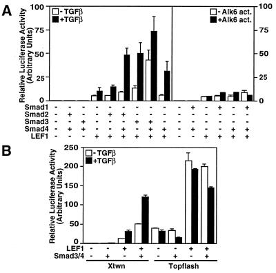
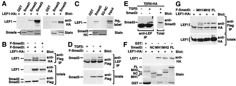
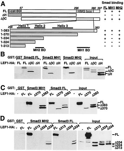
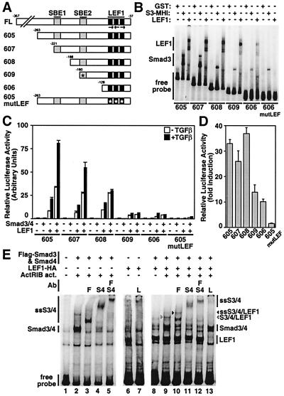
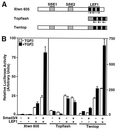
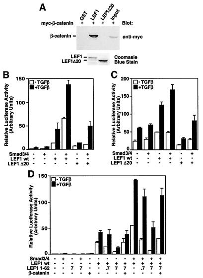
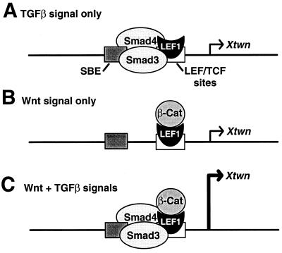
The transforming growth factor-beta (TGFbeta) and Wnt/wingless pathways play pivotal roles in tissue specification during development. Activation of Smads, the effectors of TGFbeta superfamily signals, results in Smad translocation from the cytoplasm into the nucleus where they act as transcriptional comodulators to regulate target gene expression. Wnt/wingless signals are mediated by the DNA-binding HMG box transcription factors lymphoid enhancer binding factor 1/T cell-specific factor (LEF1/TCF) and their coactivator beta-catenin. Herein, we show that Smad3 physically interacts with the HMG box domain of LEF1 and that TGFbeta and Wnt pathways synergize to activate transcription of the Xenopus homeobox gene twin (Xtwn). Disruption of specific Smad and LEF1/TCF DNA-binding sites in the promoter abrogates synergistic activation of the promoter. Consistent with this observation, introduction of Smad sites into a TGFbeta-insensitive LEF1/TCF target gene confers cooperative TGFbeta and Wnt responsiveness to the promoter. Furthermore, we demonstrate that TGFbeta-dependent activation of LEF1/TCF target genes can occur in the absence of beta-catenin binding to LEF1/TCF and requires both Smad and LEF1/TCF DNA-binding sites in the Xtwn promoter. Thus, our results show that TGFbeta and Wnt signaling pathways can independently or cooperatively regulate LEF1/TCF target genes and suggest a model for how these pathways can synergistically activate target genes.


Figures

Figure 1
Figure 1
TGFβ and Wnt signaling pathways activate Xtwn but not Topflash reporters. HepG2 cells were transfected with Xtwn-Lux (A and B) or Topflash (B) reporters alone or together with LEF1 and Smads. Cells were incubated overnight with or without 100 pM TGFβ (A Left and B) or were cotransfected with the constitutively active BMP type I receptor ALK6 (A Right). Luciferase activity was normalized to β-galactosidase activity and is expressed as the mean ± SD.
Figure 2
Figure 2
LEF1/TCF transcription factors associate with Smads. (A) In vitro interaction of LEF1 with bacterially expressed Smads. Lysates from LEF1-transfected COS-1 cells (A) or in vitro transcribed and translated LEF1 (C) were incubated with bacterially expressed GST–Smad fusion proteins, and bound material was detected by immunoblotting. Levels of GST fusion proteins were confirmed by SDS/PAGE and Coomassie blue staining (stain). (B) Interaction of LEF1 with Smads in mammalian cells. COS-1 cells were transfected with constitutively activated ActRIB, Flag-tagged Smad2, or Smad3 and HA-tagged LEF1. Cell lysates were subjected to anti-Flag antibody immunoprecipitation and were analyzed by anti-HA immunoblotting. Protein levels were determined by anti-Flag or anti-HA immunoblotting of total cell lysates. (D) TGFβ-dependent enhancement of the interaction of Smad3 with endogenous LEF1/TCF. HEK 293T cells were transfected with Flag–Smad3, Smad4, and TβRII, and cells were incubated overnight in the presence or absence of TGFβ. Endogenous LEF1/TCF was collected by immunoprecipitation with anti-LEF1/TCF antibodies, and associated Smad3 was detected by anti-Flag immunoblotting. Smad3 protein levels were determined by anti-Flag immunoblotting of total cell lysates. (E) TGFβ-dependent association of endogenous Smad3 and LEF1/TCF. HEK 293T cells, transfected with TβRII, were incubated overnight with or without TGFβ. Endogenous LEF1/TCF was collected by anti-LEF1/TCF immunoprecipitation, and associated Smad3 was detected by immunoblotting with anti-Smad2/3 antibodies. In parallel, cells were transfected with untagged Smad3, and an aliquot of total cell extracts was subjected to anti-Smad2/3 immunoblotting to confirm comigration with endogenous Smad3. (F) In vitro interaction of LEF1 with bacterially expressed FL, MH1, MH2, or nonconserved domains of Smad3. Association of LEF1 with the various Smad3 domains was conducted as in A. (G) Interaction of LEF1 with FL, MH1, or MH2 domains of Smad3 in mammalian cells. Association of LEF1 with the various Smad3 domains was conducted as in B.
Figure 3
Figure 3
Determination of the domains in LEF1 that mediate association with Smad3. (A) A schematic representation of mutant versions of LEF1 are shown. The β-catenin binding domain (β-cat BD) and the HMG box are marked. The location of the three helices within the HMG box (overline) and the MH1- and MH2-binding domains (BD; underline) are indicated. A summary of the interaction of LEF1 with FL, MH1, and MH2 domains of Smad3 is shown (right). (B–D) Interaction of wild-type or mutant LEF1 with bacterially expressed FL, MH1, or MH2 domains of Smad3. COS-1 cells were transfected with wild-type or mutant versions of LEF1 and cell lysates incubated with bacterially expressed GST fusion proteins. The associated LEF1 was visualized by anti-HA immunoblotting. Total LEF1 protein expression was confirmed by immunoblotting (input).
Figure 4
Figure 4
The SBEs and the LEF1/TCF-binding sites are required for synergistic activation of Xtwn by TGFβ and Wnt signaling pathways. (A) A schematic representation of the Xtwn promoter and the 5′ end deletion constructs with the location of the SBEs, the triple LEF1/TCF-binding sites (LEF), and the presence of point mutations (asterisk) is shown. (B) EMSA analysis of Xtwn promoter deletions. Bacterially expressed GST–LEF1, GST–Smad3 MH1 domain, or GST control were subjected to gel shift assays with 32P-labeled Xtwn promoter probes corresponding to the indicated DNA fragments. (C) HepG2 cells were transfected with Xtwn–Lux reporter constructs containing various promoter fragments alone or together with combinations of LEF1, Smad3, and Smad4 as in Fig. 1. (D) LEF1-dependent activation of luciferase activity. Luciferase activity from C is replotted as fold induction for transfectants expressing LEF1 in the absence of TGFβ treatment compared with controls transfected with Xtwn reporters alone. (E) Smads and LEF1 expressed in mammalian cells bind to the Xtwn promoter. Extracts from COS-1 cells transfected with combinations of Smad3, Smad4, LEF1, and the constitutively active activin type I receptor ActRIB were tested for DNA-binding activity on the 322-bp Xtwn promoter as in B. For supershifting assays, anti-Flag (F), anti-Smad4 (S4), or anti-LEF1/TCF (L) antibodies (Ab) were added. The migration of DNA binding and supershifted (SS) complexes are indicated. The complex containing Smad3, Smad4, and LEF1 is marked (arrow).
Figure 5
Figure 5
Introduction of SBEs into Topflash confers TGFβ-responsiveness to the promoter. (A) A schematic representation of the Xtwn, Topflash, and Twntop promoters is shown. (B) HepG2 cells were transfected with Xtwn–Lux, Topflash, or Twntop reporters alone or together with various combinations of LEF1, Smad3, and Smad4 as in Fig. 1.
Figure 6
Figure 6
TGFβ-dependent activation of LEF1 target genes occurs independently of β-catenin. (A) In vitro interaction of bacterially expressed LEF1 and LEF1 Δ20 with β-catenin. Cell lysates from COS-1 cells transfected with β-catenin were incubated with bacterially expressed GST fusion proteins of LEF1, LEF1 Δ20, or control. Bound material was visualized by anti-myc immunoblotting. (B and C) Smad enhances LEF1-dependent signaling in the absence of β-catenin. HepG2 cells were transfected with Xtwn–Lux (B) or Twntop (C) reporter alone or with Smad3, Smad4, wild-type LEF1, or LEF1 Δ20. (D) Overexpression of the β-catenin-binding domain of LEF1 does not disrupt Smad-dependent enhancement of LEF1-dependent signaling. HepG2 cells were transfected with Xtwn–Lux reporter alone or with Smad3, Smad4, wild-type LEF1, LEF1 1–62, or β-catenin. For LEF1 1–62 the amount of DNA (ng per well) is indicated.
Figure 7
Figure 7
A model for activation of specific target genes by TGFβ and Wnt pathways. Promoters with SBEs adjacent to the LEF1/TCF-binding sites can be activated by the TGFβ pathway in the absence of β-catenin (A), by Wnt signaling alone (B), or synergistically in the presence of both TGFβ and Wnt signals (C).

Similar articles

Cited by

References

    1. Cadigan K M, Nusse R. Genes Dev. 1997;11:3286–3305. - PubMed
    1. Whitman M. Genes Dev. 1998;12:2445–2462. - PubMed
    1. Massagué J. Annu Rev Biochem. 1998;67:753–791. - PubMed
    1. Wrana J L. Cell. 2000;100:189–192. - PubMed
    1. Derynck R, Zhang Y, Feng X-H. Cell. 1998;95:737–740. - PubMed

Publication types

MeSH terms

Substances