. 2000 Aug 1;97(16):8926-31.
doi: 10.1073/pnas.150238697.

Bacterial-type DNA holliday junction resolvases in eukaryotic viruses

Affiliations

Bacterial-type DNA holliday junction resolvases in eukaryotic viruses

A D Garcia et al. Proc Natl Acad Sci U S A. .

Abstract

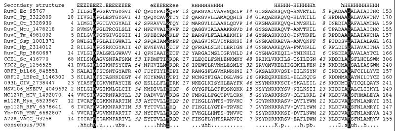
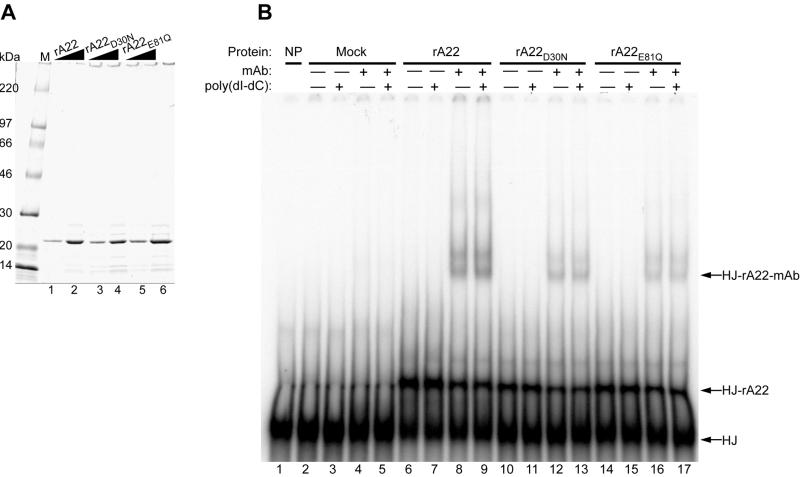
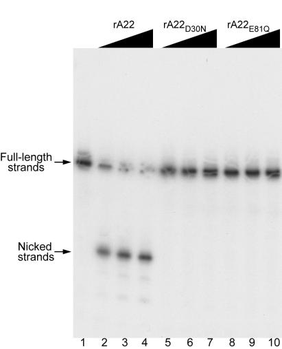
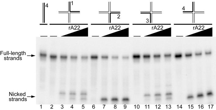
Homologous DNA recombination promotes genetic diversity and the maintenance of genome integrity, yet no enzymes with specificity for the Holliday junction (HJ)-a key DNA recombination intermediate-have been purified and characterized from metazoa or their viruses. Here we identify critical structural elements of RuvC, a bacterial HJ resolvase, in uncharacterized open reading frames from poxviruses and an iridovirus. The putative vaccinia virus resolvase was expressed as a recombinant protein, affinity purified, and shown to specifically bind and cleave a synthetic HJ to yield nicked duplex molecules. Mutation of either of two conserved acidic amino acids abrogated the catalytic activity of the A22R protein without affecting HJ binding. The presence of bacterial-type enzymes in metazoan viruses raises evolutionary questions.


Figures

Figure 1
Figure 1
Multiple sequence alignment of the RuvC family of HJ resolvases. The alignment was constructed by using the macaw program; only the five conserved motifs are shown. The lengths of the poorly conserved spacers between the motifs are indicated by italic numbers. The positions of the first and the last of the aligned amino acid residues in each sequence are also indicated. The protein designation consist of the gene, an abbreviated species name, and the GenBank identification number (separated by underlines). The consensus derived using 90% conservation is shown underneath the alignment; b indicates “big” residues (E, K, R, I, L, M, F, Y, W), h indicates hydrophobic residues (A, C, F, I, L, M, V, W, Y), s indicates small residues (A, C, S, T, D, N, V, G, P), u indicates “tiny” residues (G, A, S), and p indicates polar residues (D, E, H, K, N, Q, R, S, T). The conserved acidic residues constituting the catalytic triad of RuvC are highlighted by shading with reverse lettering. The multiple-alignment-based secondary structure prediction is shown on top of the alignment; E (e) indicates extended conformation (β-strand), and H (h) indicates α-helix (uppercase indicates the most confident prediction). Species abbreviations: Bacteria: Ct, Chlamydia trachomatis; Ec, E. coli; Hp, Helicobacter pylori; Mtu, Mycobacterium tuberculosis; Rp, Rickettsia prowazekii; Ssp, Synechocystis sp.; Tm, Thermotoga maritima; Tp, Treponema pallidum. Eukaryotic mitochondrial: Sc, Sac. cerevisiae; Sp, Sch. pombe. Viruses: biL66, LBPc2, Lactococcus lactis bacteriophages; CIV, Chilo iridescent virus; MCV, molluscum contagiosum virus; MSEPV, Melanoplus sanguinipes entomopoxvirus; Myx, myxoma virus; RFV, rabbit fibroma virus; VACC, vaccinia virus; YMV, Yaba monkey virus.
Figure 2
Figure 2
Binding of affinity-purified rA22, rA22D30N, and rA22E81Q to a synthetic HJ. (A) Recombinant polyhistidine-tagged rA22, rA22D30N, and rA22E81Q were expressed in E. coli and purified by chromatography on a metal-affinity resin, analyzed on an SDS/4–20% polyacrylamide gel, and stained with Coomassie blue. Lanes: M, molecular mass markers; 1, 2.1 μg of rA22; 2, 4.2 μg of rA22; 3, 2.3 μg of rA22D30N; 4, 4.6 μg of rA22D30N; 5, 2.6 μg of rA22E81Q; and 6, 5.2 μg of rA22E81Q. Wedges indicate increasing amounts of protein. (B) Binding of rA22, rA22D30N, and rA22E81Q to the HJ. Affinity-purified recombinant proteins or mock-affinity-purified proteins from bacterial extracts were incubated with 0.1 pmol of HJ in the presence of EDTA, and with (+) or without (−) 75-fold excess poly(dI-dC)⋅poly(dI-dC) or anti-tetrahistidine monoclonal antibody (mAb). The native products were analyzed on a 4% polyacrylamide gel. Lanes: 1, no protein; 2–5, mock-affinity-purified proteins; 6–9, 0.42 μg of rA22; 10–13, 0.46 μg of rA22D30N; 14–17, 0.52 μg of rA22E81Q. Free HJ, HJ-rA22, and HJ-rA22-mAb complexes are indicated on the right side of the autoradiogram.
Figure 3
Figure 3
Nicking of a synthetic HJ by rA22. Recombinant proteins were incubated for 30 min at 37°C with 0.2 pmol of HJ that was 5′-32P-end-labeled on strand 4. Reactions were stopped by addition of SDS and EDTA, and the products were analyzed by electrophoresis on a denaturing 12% polyacrylamide gel and visualized by autoradiography. Lanes: 1, no protein; 2–4, 0.30 to 3.0 μg of rA22; 5–7, 0.43 to 4.3 μg of rA22D30N; 8–10, 0.43 to 4.3 μg of rA22E81Q.
Figure 4
Figure 4
Substrate specificity of rA22. rA22 was incubated for 25 min at 37°C with 0.20 pmol of single-stranded, duplex, Y-junction, three-stranded junction, or four-stranded HJ that was 5′-32P-end-labeled on one strand. Reactions were stopped by addition of SDS and EDTA, and the products were analyzed by electrophoresis on a nondenaturing 10% polyacrylamide gel and visualized by autoradiography. Lanes: 1–5, no recombinant protein; 6–10, 3.2 μg of rA22. DNA substrates are shown diagrammatically above the autoradiograph with the 32P-end-labeled strand indicated in boldface.
Figure 5
Figure 5
DNA strands of HJ are nicked symmetrically by rA22. A set of four HJs, each with a different 5′-32P-end-labeled strand indicated in bold (0.1 to 0.14 pmol) was incubated with rA22 for 30 min and analyzed by electrophoresis on a denaturing polyacrylamide gel as in Fig. 3. Lanes 1, 2, 6, 10, and 14 show the mobilities of duplex or HJ probes without added proteins. The wedges above the other lanes indicate 0.34 to 3.4 μg of rA22 added to the reaction mixtures. Upper and lower arrows point to full-length and nicked strands, respectively.

Comment in

  • Resolving the relationships of resolving enzymes.
    Lilley DM, White MF. Lilley DM, et al. Proc Natl Acad Sci U S A. 2000 Aug 15;97(17):9351-3. doi: 10.1073/pnas.97.17.9351. Proc Natl Acad Sci U S A. 2000. PMID: 10944205 Free PMC article. No abstract available.

References

    1. Holliday R. Genet Res. 1964;5:282–304.
    1. Ortiz-Lombardia M, Gonzalez A, Eritja R, Aymami J, Azorin F, Coll M. Nat Struct Biol. 1999;6:913–917. - PubMed
    1. Dunderdale H J, Benson F E, Parsons C A, Sharples G J, Lloyd R G, West S C. Nature (London) 1991;354:506–510. - PubMed
    1. Iwasaki H, Takahagi M, Shiba T, Nakata A, Shinagawa H. EMBO J. 1991;10:4381–4389. - PMC - PubMed
    1. Connolly B, Parsons C A, Benson F E, Dunderdale H J, Sharples G J, Lloyd R G, West S C. Proc Natl Acad Sci USA. 1991;88:6063–6067. - PMC - PubMed

MeSH terms

LinkOut - more resources