. 2000 Jul 18;97(15):8606-11.
doi: 10.1073/pnas.150241097.

Dopamine D1 and adenosine A1 receptors form functionally interacting heteromeric complexes

Affiliations

Dopamine D1 and adenosine A1 receptors form functionally interacting heteromeric complexes

S Ginés et al. Proc Natl Acad Sci U S A. .

Abstract

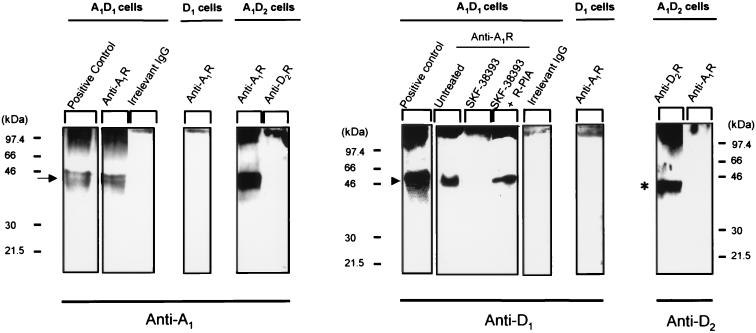
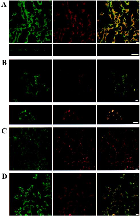
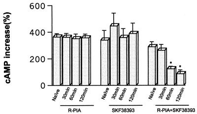
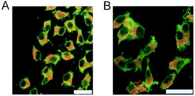
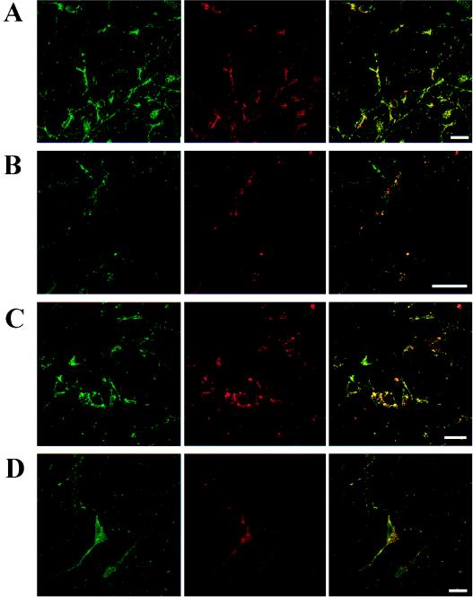
The possible molecular basis for the previously described antagonistic interactions between adenosine A(1) receptors (A(1)R) and dopamine D(1) receptors (D(1)R) in the brain have been studied in mouse fibroblast Ltk(-) cells cotransfected with human A(1)R and D(1)R cDNAs or with human A(1)R and dopamine D(2) receptor (long-form) (D(2)R) cDNAs and in cortical neurons in culture. A(1)R and D(1)R, but not A(1)R and D(2)R, were found to coimmunoprecipitate in cotransfected fibroblasts. This selective A(1)R/D(1)R heteromerization disappeared after pretreatment with the D(1)R agonist, but not after combined pretreatment with D(1)R and A(1)R agonists. A high degree of A(1)R and D(1)R colocalization, demonstrated in double immunofluorescence experiments with confocal laser microscopy, was found in both cotransfected fibroblast cells and cortical neurons in culture. On the other hand, a low degree of A(1)R and D(2)R colocalization was observed in cotransfected fibroblasts. Pretreatment with the A(1)R agonist caused coclustering (coaggregation) of A(1)R and D(1)R, which was blocked by combined pretreatment with the D(1)R and A(1)R agonists in both fibroblast cells and in cortical neurons in culture. Combined pretreatment with D(1)R and A(1)R agonists, but not with either one alone, substantially reduced the D(1)R agonist-induced accumulation of cAMP. The A(1)R/D(1)R heteromerization may be one molecular basis for the demonstrated antagonistic modulation of A(1)R of D(1)R receptor signaling in the brain. The persistence of A(1)R/D(1)R heteromerization seems to be essential for the blockade of A(1)R agonist-induced A(1)R/D(1)R coclustering and for the desensitization of the D(1)R agonist-induced cAMP accumulation seen on combined pretreatment with D(1)R and A(1)R agonists, which indicates a potential role of A(1)R/D(1)R heteromers also in desensitization mechanisms and receptor trafficking.


Figures

Figure 1
Figure 1
Coimmunoprecipitation of A1R and D1R. Cell membranes from A1/D1, A1/D2, or D1 cells were obtained and processed for immunoprecipitation (see Methods) by using the purified anti-A1R antibody PC11, the anti-D2R antibody, or an irrelevant goat IgG; all were covalently coupled to protein A-Sepharose. Immunoblottings of cell lysates (positive control) and immunoprecipitates were performed to detect A1R with anti-A1R antibody, D1R with anti-D1R antibody, or D2R with anti-D2R antibody. When indicated, A1/D1-cotransfected cells were incubated for 1 h with 10 μM SKF-38393 in the absence or presence of 100 nM R-PIA. The arrow indicates the band for A1R, the arrowhead the band corresponding to D1R, and the asterisk the band for D2R.
Figure 2
Figure 2
Distribution of A1R and D1R in A1/D1-cotransfected fibroblast cells. Cells were incubated for 1 h with medium in the absence (A) or presence of 100 nM R-PIA (B), 10 μM SKF-38393 (C), or 100 nM R-PIA plus 10 μM SKF-38393 (D) and were processed for immunostaining (see Methods) by using fluorescein (green)-conjugated rabbit anti-A1R antibody and a Texas red-conjugated rabbit anti-D1R antibody. The cells were analyzed by confocal laser microscopy. Superimposition of images (Right images in each panel) reveals the colocalization of A1R and D1R in yellow. (A Lower) A vertical section of representative cells is also shown. (B Lower) A magnification of a representative cell is also given. (Scale bars: 10 μm.)
Figure 3
Figure 3
cAMP accumulation induced by incubation with 10 μM SKF-38393 (15 min) after pretreatment of A1/D1-cotransfected cells with 100 nM R-PIA and/or 10 μM SKF-38393. Control cells (naive) were treated with medium alone for 120 min. Data represent the means ± SEM (n = 6) of the percentage of increase versus basal values. The basal values of cAMP for the groups treated with R-PIA, SKF-38393, and R-PIA plus SKF-38393 were (means ± SEM, in pmol/mg of protein; n = 4) 11,020 ± 1,345; 24,060 ± 2,279; and 14,030 ± 1,375; respectively. Repeated measures ANOVA with post hoc Scheffé's test: *, P < 0.01 with respect to control cells (not significantly different from basal values).
Figure 4
Figure 4
Distribution of A1R and D2R in A1/D2-cotransfected fibroblast cells. Cells were processed for immunostaining (see Methods) by using fluorescein (green)-conjugated rabbit anti-A1R antibody and a Texas red-conjugated rabbit anti-D2R antibody. The cells were analyzed by confocal laser microscopy. (B) Cells were treated with 100 nM R-PIA. Superimposition of images reveals the lack colocalization of A1R and D2R. (Scale bars: 10 μm.)
Figure 5
Figure 5
Distribution of A1R and D1R in primary cultures of cortical neurons. Cells were incubated for 1 h with medium in the absence (A) or presence of 100 nM R-PIA (B), 10 μM SKF-38393 (C), or 100 nM R-PIA plus 10 μM SKF-38393 (D) and were processed for immunostaining (see Methods) by using fluorescein (green)-conjugated rabbit anti-A1R antibody and a Texas red-conjugated rabbit anti-D1R antibody. The cells were analyzed by confocal microscopy. Superimposition of images (Right images in each panel) reveals the colocalization of A1R and D1R in yellow. (Scale bars: 10 μm.)

References

    1. Agnati L F, Fuxe K, Benfenati F, Celani M F, Battistini N, Mutt V, Cavicchioli L, Galli G, Hokfelt T. Neurosci Lett. 1983;35:179–183. - PubMed
    1. Fuxe K, Agnati L F, editors. Receptor-Receptor Interactions. Wenner-Gren Center Interactional Symposium Series. New York: Macmillan; 1987.
    1. Zoli M, Agnati L F, Hedlund P B, Li X M, Ferré S, Fuxe K. Mol Neurobiol. 1993;7:293–334. - PubMed
    1. Ng G Y, George S R, Zastawny R L, Caron M, Bouvier M, Dennis M, O'Dowd B F. Biochemistry. 1993;32:11727–11733. - PubMed
    1. Ng G Y, O'Dowd B F, Caron M, Dennis M, Brann M R, George S R. J Neurochem. 1994;63:1589–1595. - PubMed

Publication types

MeSH terms

LinkOut - more resources