. 2000 Aug;20(15):5381-91.
doi: 10.1128/MCB.20.15.5381-5391.2000.

Role of the IkappaB kinase complex in oncogenic Ras- and Raf-mediated transformation of rat liver epithelial cells

Affiliations

Role of the IkappaB kinase complex in oncogenic Ras- and Raf-mediated transformation of rat liver epithelial cells

M Arsura et al. Mol Cell Biol. 2000 Aug.

Abstract

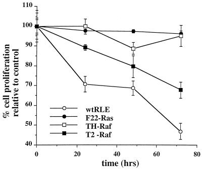
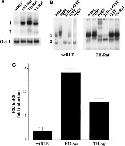
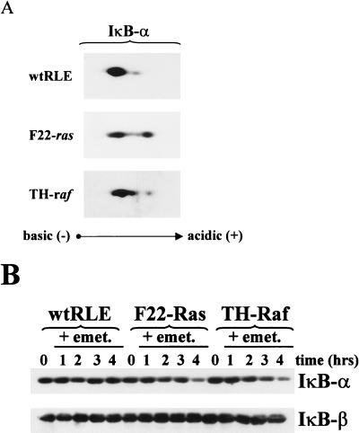
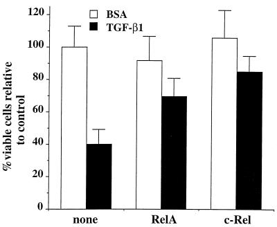
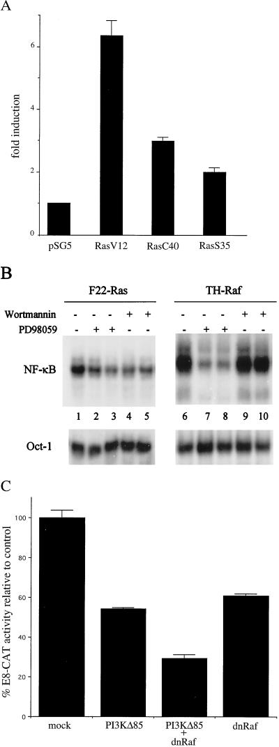
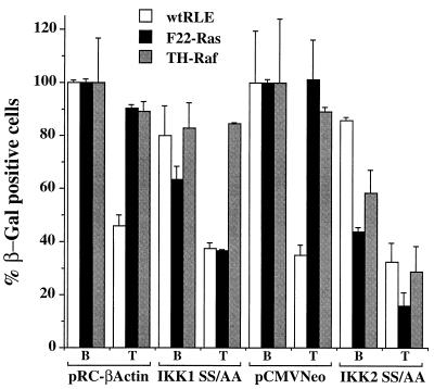
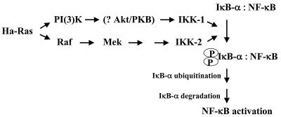
NF-kappaB/Rel factors have been implicated in the regulation of liver cell death during development, after partial hepatectomy, and in hepatocytes in culture. Rat liver epithelial cells (RLEs) display many biochemical and ultrastructural characteristics of oval cells, which are multipotent cells that can differentiate into mature hepatocytes. While untransformed RLEs undergo growth arrest and apoptosis in response to transforming growth factor beta1 (TGF-beta1) treatment, oncogenic Ras- or Raf-transformed RLEs are insensitive to TGF-beta1-mediated growth arrest. Here we have tested the hypothesis that Ras- or Raf-transformed RLEs have altered NF-kappaB regulation, leading to this resistance to TGF-beta1. We show that classical NF-kappaB is aberrantly activated in Ras- or Raf-transformed RLEs, due to increased phosphorylation and degradation of IkappaB-alpha protein. Inhibition of NF-kappaB activity with a dominant negative form of IkappaB-alpha restored TGF-beta1-mediated cell killing of transformed RLEs. IKK activity mediates this hyperphosphorylation of IkappaB-alpha protein. As judged by kinase assays and transfection of dominant negative IKK-1 and IKK-2 expression vectors, NF-kappaB activation by Ras appeared to be mediated by both IKK-1 and IKK-2, while Raf-induced NF-kappaB activation was mediated by IKK-2. NF-kappaB activation in the Ras-transformed cells was mediated by both the Raf and phosphatidylinositol 3-kinase pathways, while in the Raf-transformed cells, NF-kappaB induction was mediated by the mitogen-activated protein kinase cascade. Last, inhibition of either IKK-1 or IKK-2 reduced focus-forming activity in Ras-transformed RLEs. Overall, these studies elucidate a mechanism that contributes to the process of transformation of liver cells by oncogene Ras and Raf through the IkappaB kinase complex leading to constitutive activation of NF-kappaB.


Figures

FIG. 1
FIG. 1
Oncogenic Ras- or Raf-transformed RLEs are resistant to TGF-β1-induced cell growth arrest. Cultures of wt and F22-Ras, TH-Raf, and T2-Raf RLEs were incubated in medium containing TGF-β1 (5 ng/ml) or BSA carrier solution for 24, 48, and 72 h. Cell proliferation was monitored by conversion of MTS to its formazan product. Means and standard deviations are representative of two independent experiments carried out in triplicate.
FIG. 2
FIG. 2
NF-κB is aberrantly expressed in Ras- or Raf-transformed RLEs. (A) Ras- and Raf-transformed cells display elevated levels of NF-κB binding. To measure the levels of NF-κB binding activity in wt and transformed RLEs, EMSA was performed using URE-κB motif from the c-myc gene as a probe (20) and nuclear extracts from exponentially growing wt and transformed (F-22, TH, and T2) RLEs. As control for equal loading, EMSA was also performed with an Oct-1 probe. (B) Transformed cells express classical NF-κB. For supershift analysis, following a 30-min incubation of nuclear extracts from the wt RLE or TH-Raf cells with the UREκB probe, 1 μl of antibody against either the p50 (SC114), p65 (sc-109), or c-Rel (cross-reacting v-Rel antibody, kindly provided by N. Rice) protein was added as indicated. The reaction mixture was incubated for an additional 1 h and subjected to EMSA. Alternatively, 100 ng of either IκB-α–GST or GST protein was added to the reaction mixture. (C) Transformed RLEs display elevated NF-κB activity. Wild-type and transformed RLEs in P60 dishes were transiently transfected by lipofection, in duplicate, with 6 μg of E8 or dmE8 reporter construct. The E8 vector has two copies of the URE-κB motif in front of the TK promoter driving the CAT reporter; the double-mutant vector has two copies of a mutant version of the URE with two G-to-C conversions, which is unresponsive to NF-κB transactivation (dmE8). The values for E8 CAT activity are represented as fold induction over dmE8 CAT activity, which was set at 1.0 for each cell line.
FIG. 3
FIG. 3
IκB-α protein is more phosphorylated and has a shorter half-life in F22-Ras and TH-Raf cells than in wt RLEs. (A) Phosphorylation state. Cytoplasmic extracts (40 μg) from exponentially growing wt, F22-Ras and TH-Raf RLEs, treated for 1 h with 40 μM calpain inhibitor I to inhibit IκB-α degradation, were resolved on isoelectric focusing gels, separated according to molecular weight by SDS-PAGE, and subjected to immunoblot analysis using an antibody preparation raised against the IκB-α product (SC-371). (B) Half-life of decay. Exponentially growing RLEs were treated with the protein synthesis inhibitor emetine (emet; 10 μg/ml) for 1 to 4 h. Cytoplasmic extracts (20 μg) were then subjected to immunoblot analysis for IκB-α as described above.
FIG. 4
FIG. 4
F22, TH, and T2 cells maintain NF-κB DNA binding activity following TGF-β1 treatment. Wild-type and transformed cells were treated for 24 h in the presence of TGF-β1 (5 ng/ml; T) or BSA carrier solution (B) as a control, and the levels of NF-κB binding activity to the URE-κB probe were monitored by EMSA analysis for Fig. 2B above.
FIG. 5
FIG. 5
NF-κB/Rel factors rescue wt RLEs from TGF-β1-mediated apoptosis. RLEs were plated at 70% confluence in 96-well plates and transiently transfected by lipofection. DNAs transfected included 25 ng of β-Gal-expressing vector pON407, in which the five putative NF-κB sites within the CMV promoter were removed in the presence of 75 ng of vector directing expression of either the RelA (p65) (11) or c-Rel (1) subunit or pUC18 DNA as control (none). Six hours after transfection, TGF-β1 (5 ng/ml) or BSA carrier solution was added, and cells were incubated for an additional 48 h. Cells were stained with X-Gal as described previously (9), and the viable (blue) cells were counted. Values are given relative to that for control cells (transfected with pUC18 DNA and treated with BSA), which was set at 100%. Means and standard deviations are representative of two independent experiments carried out in triplicate.
FIG. 6
FIG. 6
Inhibition of NF-κB activity restores TGF-β1-mediated cell killing of oncogenic Ras- or Raf-transformed RLEs. F22-Ras (A) or TH-Raf (B) cells were lipofected with 25 ng of β-Gal-expressing vector pON407 in the presence of either 75 ng of pMT2T-IκB-α construct (11), directing expression of the inhibitor IκB-α, or pUC18 DNA, as indicated. Six hours after transfection, TGF-β1 (5 ng/ml) or BSA carrier solution was added, and cells were incubated for an additional 48 h. Alternatively, cells were cotransfected with 25 ng of pON407 plus 37.5 ng of vector expressing RelA in the absence or presence of 37.5 ng of pMT2T-IκB-α or pUC18 DNA. Cells were stained with X-Gal, and the viable (blue) cells were counted. Values are given relative to that for control cells, i.e., transfected with pUC18 DNA and treated with BSA, which was set at 100%. Means and standard deviations are representative of two independent experiments carried out in triplicate.
FIG. 7
FIG. 7
IKK activity mediates constitutive NF-κB activation in oncogenic Ras- or Raf-transformed RLEs. (A) Inhibition of IKK activity. Cells were transiently transfected in P60 dishes with 2 μg of E8 or dmE8 NF-κB CAT construct in the absence or presence of 2 μg of vector directing expression dnIKK form IKK-1 SS/AA or IKK-2 SS/AA (51). As controls, the parental pRC-βactin and pCMV-Neo constructs, respectively, were similarly transfected. The values for E8 CAT activity are represented as fold induction over dmE8 CAT activity, which was set at 1.0 for each cell line. (B) Mutant version of IKKAP1 inhibits NF-κB activation in F22-Ras cells. Cells in P60 dishes were transiently transfected with 2 μg of E8 or dmE8 NF-κB CAT construct in the absence or presence of 2 μg of DNA of a vector directing expression of either full-length (FL) IKKAP1 or its C-terminus-deleted version ΔC IKKAP1 (51). In addition, 1 μg of simian virus 40–β-Gal expression vector was added to normalize for transfection efficiency. As control, the parental pCDNA3-EE construct was similarly transfected. The values are represented as percentage relative to the CAT activity of the E8 reporter in the presence of the parental vector, which was set at 100%. Means and standard deviations are representative of two independent experiments carried out in duplicate. (C) Kinase assays. (Top) Cells were transiently transfected with 5 μg of vector directing expression of wt IKK-1 or IKK-2 as described above. Following immunoprecipitation with antibodies against IKK-1 or IKK-2, extracts (10 μg) were subjected to kinase assays using either wt IκB-α–GST or the Ser32/36 double-mutant IκB-α–GST version, which cannot be phosphorylated. (Bottom) Equal aliquots of the immunoprecipitates from the lanes in the top panel, as indicated, were subjected to immunoblotting for IKK-1 and IKK-2 protein. (D) Extracts from exponentially growing cells (80 μg) were immunoprecipitated with antibodies against IKK-1 or IKK-2. Immunoprecipitated proteins were subjected to kinase assay using wt IκB-α–GST as substrate.
FIG. 8
FIG. 8
Oncogenic Ras induces NF-κB translocation through both the PI(3)K and Raf pathways. (A) Wild-type RLE cells were transiently transfected in P100 dishes with 3 μg of wt E8 NF-κB CAT construct in the absence or presence of 1 μg of pSG5-V12 H-ras, pSG5-V12C40 H-ras, and pSG5-V12S35 H-ras expression vectors. As a control, the parental pSG5 vector was similarly transfected. The final amount of DNA was adjusted to 12 μg using pSG5 DNA. The values for E8 CAT activity are represented as fold induction over the E8 CAT activity in cells transfected with the pSG5 vector alone, which was set at 1. (B) F22-Ras and TH-Raf cells were treated for 3 h in the presence of 100 (lanes 2 and 3) or 150 (lanes 7 and 8) μM PD98059. Alternatively, F22-Ras and TH-Raf cells were treated for 3 h with 10 (lanes 4 and 5) or 100 (lanes 9 and 10) wortmannin. As a control, cells were treated with carrier dimethyl sulfoxide alone (lanes 1 and 6). Nuclear extracts were prepared, and the levels of binding activity to the URE (NF-κB) and Oct-1 probes were monitored by EMSA. (C) F22-Ras cells were transiently transfected in P100 dishes with 3 μg of wt E8 NF-κB CAT construct with 1 μg of SR-αΔp85 (PI3KD85), 1 μg of pUC19-LTR-ΔRaf (dnRaf), or both. As controls, the parental vectors were similarly transfected to bring the final amount of DNA to 12 μg. The values for E8 CAT activity are expressed as percentage of the E8 CAT activity in cells transfected with the parental vectors alone, which was set at 100.
FIG. 9
FIG. 9
Inhibition of IKK activity renders transformed RLEs sensitive to TGF-β1-induced apoptosis. Normal and transformed RLEs were lipofected with 25 ng of β-Gal-expressing vector pON407 in the presence of either 25 ng of IKK-1 SS/AA or IKK-2 SS/AA expression vector DNA. As controls, the respective parental pRC-βactin and pCMV-Neo constructs were similarly transfected. Six hours after transfection, TGF-β1 (5 ng/ml) or BSA carrier solution was added to the culture medium, and the cells were incubated for an additional 48 h. Viable cells and values were determined as described in the legend to Fig. 6.
FIG. 10
FIG. 10
Inhibition of IKK-1 or IKK-2 activity reduces oncogenic Ras focus-forming activity in RLEs. F22-Ras cells were lipofected as described in the legend to Fig. 9. After 24 h, cells were plated in soft agar; after 2 weeks, the number of foci was scored. Means and standard deviation are representative of two independent experiments carried out in duplicate.
FIG. 11
FIG. 11
Schematic representation of Ha-Ras-mediated activation of NF-κB through IKK complex activation. Ras leads to activation of NF-κB via two pathways: PI(3)K and Raf, as discussed in the text. Akt/PKB, protein kinase B.

References

    1. Arsura M, FitzGerald M J, Fausto N, Sonenshein G E. NF-κB/Rel blocks transforming growth factor-β1-induced apoptosis of murine hepatocyte cell lines. Cell Growth Differ. 1997;8:1049–1059. - PubMed
    1. Arsura M, Wu M, Sonenshein G E. TGF-β1 inhibits NF-κB/Rel activity inducing apoptosis of B cells: transcriptional activation of IκB-α. Immunity. 1996;5:31–40. - PubMed
    1. Baeuerle P A, Henkel T. Function and activation of NF-κB in the immune system. Annu Rev Immunol. 1994;12:141–179. - PubMed
    1. Baldwin A S., Jr The NF-κB and IκB proteins: new discoveries and insights. Annu Rev Immunol. 1996;14:649–681. - PubMed
    1. Ballard D, Dixon E, Peffer N, Bogard H, Doerre S, Stein B, Greene W. The 65-kDa subunit of human NF-κB functions as a potent transcriptional activator and a target for v-Rel-mediated repression. Proc Natl Acad Sci USA. 1992;85:1875–1880. - PMC - PubMed

Publication types

MeSH terms