. 2000 Aug;20(15):5503-15.
doi: 10.1128/MCB.20.15.5503-5515.2000.

CD40 induces apoptosis in carcinoma cells through activation of cytotoxic ligands of the tumor necrosis factor superfamily

Affiliations

CD40 induces apoptosis in carcinoma cells through activation of cytotoxic ligands of the tumor necrosis factor superfamily

A G Eliopoulos et al. Mol Cell Biol. 2000 Aug.

Abstract

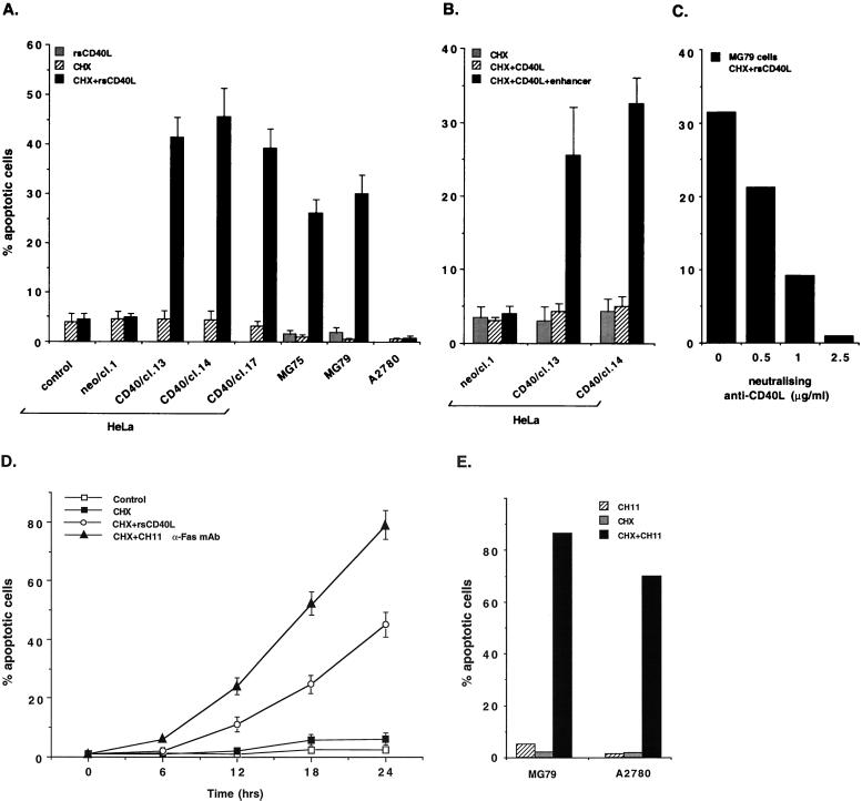
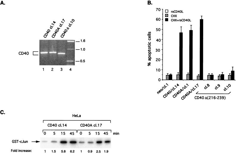
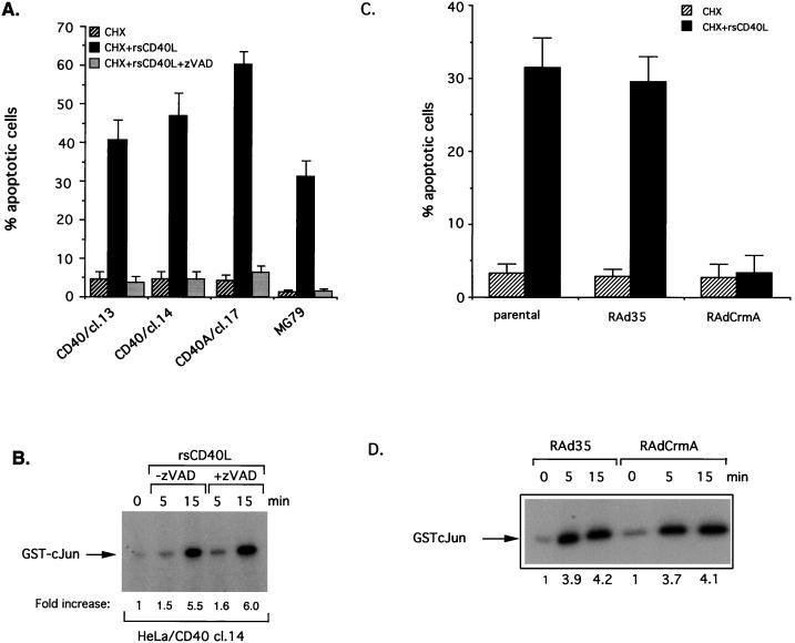
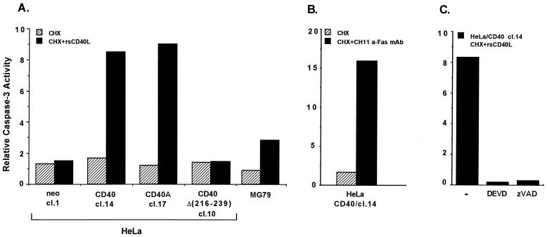
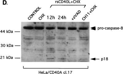
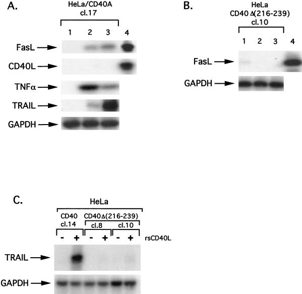
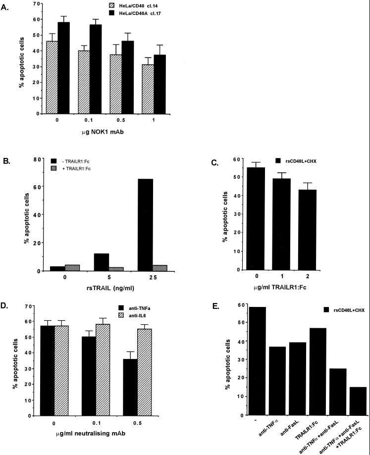
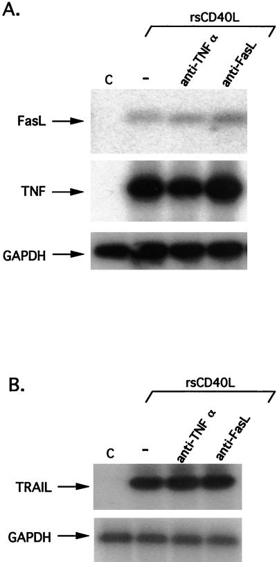
CD40, a tumor necrosis factor (TNF) receptor (TNFR) family member, conveys signals regulating diverse cellular responses, ranging from proliferation and differentiation to growth suppression and cell death. The ability of CD40 to mediate apoptosis in carcinoma cells is intriguing given the fact that the CD40 cytoplasmic C terminus lacks a death domain homology with the cytotoxic members of the TNFR superfamily, such as Fas, TNFR1, and TNF-related apoptosis-inducing ligand (TRAIL) receptors. In this study, we have probed the mechanism by which CD40 transduces death signals. Using a trimeric recombinant soluble CD40 ligand to activate CD40, we have found that this phenomenon critically depends on the membrane proximal domain (amino acids 216 to 239) but not the TNFR-associated factor-interacting PXQXT motif in the CD40 cytoplasmic tail. CD40-mediated cytotoxicity is blocked by caspase inhibitors, such as zVAD-fmk and crmA, and involves activation of caspase 8 and caspase 3. Interestingly, CD40 ligation was found to induce functional Fas ligand, TRAIL (Apo-2L) and TNF in apoptosis-susceptible carcinoma cells and to up-regulate expression of Fas. These findings identify a novel proapoptotic mechanism which is induced by CD40 in carcinoma cells and depends on the endogenous production of cytotoxic cytokines and autocrine or paracrine induction of cell death.


Figures

FIG. 1
FIG. 1
Recombinant soluble forms of CD40L induce apoptosis in carcinoma cells. (A) An rsCD40L molecule in which the extracellular domain of CD40L is linked to an isoleucine zipper trimerization motif induces cell death in carcinoma cells. The effect of CD40 ligation on survival was assessed by propidium iodide staining and fluorescence microscopy, and data are depicted as percentages of apoptotic cells (y axis) relative to untreated controls. Mean values ± standard deviations (error bars) from at least three independent experiments are shown. cl., clone. (B) CD40 oligomerization is required for effective activation of death signals in susceptible carcinoma cell lines. CD40-transfected HeLa cells were treated for 6 h with a monomeric form of CD40L (1 μg/ml; Alexis Corporation) in the presence or absence of an enhancer cross-linking molecule and then cocultured with CHX for an additional 24 h time period before apoptotic cells were counted. CD40L induced cell death only in the presence of the cross-linking antibody. No cytotoxic effect was noted upon treatment with the enhancer molecule alone (data not shown). Error bars, standard deviations. (C) Specificity of rsCD40L-induced cell death. MG79 ovarian carcinoma cells were pretreated with neutralizing anti-CD40L antibody and then exposed to rsCD40L (1 μg/ml) and CHX (8 μg/ml) treatment, as described in Materials and Methods. Apoptotic cells were counted, and mean values from two independent experiments are shown. (D) CD40 engagement induces a delayed and reduced apoptotic response compared to Fas. HeLa/CD40 clone 14 cells were treated for 6 h with rsCD40L (1 μg/ml) or CH11 (10 ng/ml) anti-Fas MAb and then cocultured with CHX for various times (0, 6, 12, 18, or 24 h). Apoptotic cells were counted and mean values ± standard deviations (error bars) from three independent experiments are shown. (E) Fas induces high levels of apoptosis in carcinoma cells which respond (MG79 ovarian carcinoma) or are resistant (A2780 ovarian carcinoma) to CD40-mediated cytotoxicity. Data shown are representative of two independent experiments.
FIG. 2
FIG. 2
A membrane-proximal domain but not a PXQXT motif in the CD40 cytoplasmic tail is critical for CD40L-induced apoptosis in carcinoma cells. (A) Expression of CD40 in representative CD40 and mutated CD40-transfected HeLa clones was verified by RT-PCR. PCR-amplified CD40 cDNA from HeLa/CD40 clone (cl.) 14 (lane 1), HeLa/CD40A cl. 17 (lane 2) and HeLa/CD40Δ(216-239) cl. 10 (lane 3) is shown. The higher electrophoretic mobility of the HeLa/CD40Δ(216-239) PCR product (lane 3) is consistent with the presence of a 23-aa deletion in the cytoplasmic tail of CD40. Lane 4 contains a molecular weight marker (labeled [in thousands] at right). (B) HeLa/CD40A but not HeLa/CD40Δ(216-239) cells are susceptible to CD40-mediated cell death. Cells were treated as described in the legend to Fig. 1A, and the percentage of apoptotic cells relative to untreated controls is depicted in histogram form. The percentage of apoptotic HeLa/CD40 cl. 14 cells is shown for comparison. Data are the mean values ± standard deviations (error bars) from three independent experiments. (C) HeLa/CD40A cells demonstrate decreased and delayed induction of JNK compared to wild-type CD40-expressing HeLa cells in response to CD40 stimulation. HeLa transfectants were treated with rsCD40L (1 μg/ml) for various time points (0, 5, 15, or 45 min), and cell lysates were subjected to immune complex kinase assays using glutathione S-transferase–cJun (aa 1 to 89) as substrate. Relative kinase activities were determined on a phosphorimager. Three independent experiments were performed and gave similar results.
FIG. 3
FIG. 3
Caspase inhibitors block CD40-induced apoptotic but not JNK signals. (A) zVAD-fmk suppresses CD40-mediated apoptosis in HeLa/CD40, HeLa/CD40A, and MG79 cells. Cells were pretreated with zVAD-fmk for 30 min and then exposed to rsCD40L for 6 h before addition of CHX. The percentage of apoptotic cells relative to untreated controls was evaluated 24 h later. Data are the mean values ± standard deviations (error bars) from three independent experiments. cl., clone. (B) Exposure of CD40-expressing cells to concentrations of zVAD-fmk which block apoptosis does not interfere with CD40-mediated JNK activation. HeLa/CD40 cl. 14 cells were pretreated for 30 min with 25 μM zVAD-fmk or left untreated and then stimulated with rsCD40L (1 μg/ml) for various time intervals before being analyzed for endogenous JNK activity. (C) CrmA expression suppresses CD40-mediated apoptosis in MG79 ovarian carcinoma cells. Cells infected with RAd-CrmA or RAd35 control virus at a multiplicity of infection of 100 were treated with rsCD40L and CHX for 24 h as described in the legend to Fig. 1A, and the percentage of apoptotic cells relative to untreated controls was evaluated. Mean values ± standard deviations (error bars) from three independent experiments are shown. (D) CrmA does not influence JNK signalling. MG79 cells infected with RAd-CrmA or RAd35 control virus at a multiplicity of infection of 100 were treated with rsCD40L (1 μg/ml) for various time intervals and then analyzed for endogenous JNK using GST-cJun(1-89) as a substrate. Relative kinase activities are shown. Values along bottom indicate fold increase of MG79.
FIG. 4
FIG. 4
CD40 ligation mediates caspase activation in apoptosis-susceptible carcinoma cells. (A) Caspase 3 activity is induced in response to rsCD40L and CHX treatment and correlates with CD40-mediated induction of cell death in carcinoma cell lines. Active caspase 3 in lysates from rsCD40L and CHX-treated cells was measured using a colorimetric caspase 3 activity kit and Ac-DEVD-pNA as the substrate. Values shown represent the relative increase in caspase 3 activity compared to that in untreated cultures, given the arbitrary value of 1, and are representative of three independent experiments. cl., clone. (B) As a positive control, treatment of HeLa/CD40 cl. 14 cells with CH11 anti-Fas MAb (10 ng/ml) and CHX (50 μg/ml) induced robust caspase 3 activity. (C) Specificity of CD40-mediated caspase 3 activity. Lysates from rsCD40L and CHX-treated HeLa/CD40 cl. 14 cells were incubated with 0.1 μM Ac-DEVD-CHO or zVAD-fmk before being analyzed for caspase 3 activity. (D) Treatment of HeLa/CD40A cl. 17 cells with rsCD40L and CHX for 12 or 24 h (lanes 3 and 4, respectively) results in caspase 8 activation, as determined by the decrease in pro-caspase 8 levels (marked with arrowheads) and the appearance of the p18 cleaved, active form (arrow) in immunoblot analysis. As a negative control, untreated cultures or cells exposed to CHX alone did not demonstrate caspase 8 activity (lanes 1 and 2), while the p18 active form was detected in lysates from cells treated for 12 h with anti-Fas (10 ng/ml) in the presence of CHX (lane 6). Pretreatment with 50 μM zVAD-fmk suppressed CD40-mediated caspase 8 activation (lane 5).
FIG. 4
FIG. 4
CD40 ligation mediates caspase activation in apoptosis-susceptible carcinoma cells. (A) Caspase 3 activity is induced in response to rsCD40L and CHX treatment and correlates with CD40-mediated induction of cell death in carcinoma cell lines. Active caspase 3 in lysates from rsCD40L and CHX-treated cells was measured using a colorimetric caspase 3 activity kit and Ac-DEVD-pNA as the substrate. Values shown represent the relative increase in caspase 3 activity compared to that in untreated cultures, given the arbitrary value of 1, and are representative of three independent experiments. cl., clone. (B) As a positive control, treatment of HeLa/CD40 cl. 14 cells with CH11 anti-Fas MAb (10 ng/ml) and CHX (50 μg/ml) induced robust caspase 3 activity. (C) Specificity of CD40-mediated caspase 3 activity. Lysates from rsCD40L and CHX-treated HeLa/CD40 cl. 14 cells were incubated with 0.1 μM Ac-DEVD-CHO or zVAD-fmk before being analyzed for caspase 3 activity. (D) Treatment of HeLa/CD40A cl. 17 cells with rsCD40L and CHX for 12 or 24 h (lanes 3 and 4, respectively) results in caspase 8 activation, as determined by the decrease in pro-caspase 8 levels (marked with arrowheads) and the appearance of the p18 cleaved, active form (arrow) in immunoblot analysis. As a negative control, untreated cultures or cells exposed to CHX alone did not demonstrate caspase 8 activity (lanes 1 and 2), while the p18 active form was detected in lysates from cells treated for 12 h with anti-Fas (10 ng/ml) in the presence of CHX (lane 6). Pretreatment with 50 μM zVAD-fmk suppressed CD40-mediated caspase 8 activation (lane 5).
FIG. 5
FIG. 5
CD40 ligation induces expression of Fas, FasL, TRAIL, and TNF in apoptosis-susceptible carcinoma cell lines. (A) CD40 ligation induces the expression of cytotoxic members of the TNF superfamily in apoptosis-susceptible carcinoma cells. RNA was isolated from HeLa/CD40A clone (cl.) 17 cells treated with rsCD40L (1 μg/ml) for 0, 2, or 12 h (lanes 1 to 3, respectively) and subjected to RT-PCR analysis for FasL, CD40L, TNF, or TRAIL (Apo-2L) expression, as described in Materials and Methods. Hybridization signals were normalized using GAPDH as the control. As a positive control for FasL and CD40L expression, RNA from RAd-FasL-infected MG79 cells (Knox et al., unpublished data) or from mouse L cells transfected with human CD40L was used (lane 4). Data are representative of at least three independent experiments. (B) HeLa/CD40Δ(216-239) cells fail to activate FasL RNA in response to CD40 ligation. RNA isolated from a representative HeLa/CD40Δ(216-239) clone treated with rsCD40L (1 μg/ml) for 0, 2, or 12 h (lanes 1 to 3, respectively) was subjected to RT-PCR analysis for expression of FasL or GAPDH. Lane 4 is a positive control for FasL expression as described above. (C) TRAIL is induced in CD40- but not CD40Δ(216-239)-transfected HeLa cells. HeLa/CD40 cl. 14, HeLa/CD40Δ(216-239) cl. 8, or HeLa/CD40Δ(216-239) cl. 10 cells were exposed to rsCD40L (1 μg/ml) for 12 h (lanes 2, 4, and 6, respectively) or left untreated (lanes 1, 3, and 5, respectively) before being analyzed for TRAIL or GAPDH expression. Two independent experiments were performed and gave similar results.
FIG. 6
FIG. 6
Inhibition of CD40-mediated FasL, TRAIL, and TNF production suppresses CD40L-induced apoptosis. (A) The neutralizing anti-FasL MAb NOK1 partially inhibits apoptosis induced by CD40L and CHX treatment of HeLa/CD40 clone (cl.) 14 and HeLa/CD40A cl. 17 cells. The percentage of apoptotic cells relative to untreated controls (mean values ± standard deviations [error bars]) from three independent experiments is shown. (B) Soluble recombinant TRAIL induces apoptosis in HeLa/CD40A cl. 17 cells when protein synthesis is inhibited. Cells were treated for 6 h with TRAIL (5 or 25 ng/ml) with enhancer in the presence (+) or absence (−) of a neutralizing TRAILR1:Fc hybrid and then incubated for 24 h in the presence of CHX, before being analyzed for cell death. Data are representative of two independent experiments. (C) A soluble TRAILR1:Fc hybrid confers only a small inhibitory effect on CD40-mediated apoptosis. HeLa/CD40A cl. 17 cells were pretreated with TRAILR1:Fc for 1 h and rsCD40L (1 μg/ml) was then added for 6 additional h. Following CHX treatment, the percentage of apoptotic cells was determined. Mean values ± standard deviations (error bars) from three independent experiments are shown. (D) A neutralizing anti-TNF but not an anti-IL-6 MAb partially inhibit CD40-mediated cell death in HeLa/CD40A cl. 17 cells. Mean values ± standard deviations (error bars) from three independent experiments are shown. Neutralizing anti-TNF MAb was also able to inhibit CD40L-induced cytotoxicity in HeLa/CD40 cl. 14 cells (data not shown). (E) Simultaneous inhibition of FasL, TNF, and TRAIL significantly suppresses CD40-mediated cell death. HeLa/CD40A cl. 17 cells were pretreated with NOK1 (1 μg/ml), anti-TNF-α (0.5 μg/ml), and TRAILR1:Fc (2 μg/ml) for 1 h, and cells were then incubated with rsCD40L (1 μg/ml) for 6 additional hours. Following CHX treatment, the percentage of apoptotic cells was determined. Data shown are representative of three independent experiments.
FIG. 7
FIG. 7
CD40 ligation independently induces the expression of FasL, TNF, and TRAIL. (A) HeLa/CD40A clone 17 cells were pretreated for 1 h with anti-TNF-α (0.5 μg/ml) (lane 3) or NOK1 (1 μg/ml) (lane 4) or were left untreated (lane 2) and then were incubated for 2 h with rsCD40L (1 μg/ml) (lanes 2 to 4) in the presence of the neutralizing reagents, before being analyzed for FasL, TNF, or GAPDH RNA levels by RT-PCR. (B) HeLa/CD40A clone 17 cells were pretreated with neutralizing reagents for 1 h as described above and then exposed to rsCD40L (1 μg/ml) for 12 h before being analyzed for TRAIL or GAPDH RNA levels by RT-PCR.

Similar articles

Cited by

References

    1. Afford S C, Randhawa S, Eliopoulos A G, Hubscher S G, Young L S, Adams D H. CD40 activation induces apoptosis in cultured human hepatocytes via induction of cell surface Fas ligand expression and amplifies Fas-mediated hepatocyte death during allograft rejection. J Exp Med. 1999;189:441–446. - PMC - PubMed
    1. Baker M P, Eliopoulos A G, Young L S, Armitage R J, Gregory C D, Gordon J. Prolonged phenotypic, functional and molecular changes in group I Burkitt lymphoma cells on short term exposure to CD40 ligand. Blood. 1998;92:2830–2843. - PubMed
    1. Barkett M, Gilmore T D. Control of apoptosis by Rel/NF-κB transcription factors. Oncogene. 1999;18:6910–6924. - PubMed
    1. Blake N, Lee S, Redchenko I, Thomas W, Steven N, Leese A, Steigerwald-Mullen P, Kurilla M G, Frappier L, Rickinson A B. Human CD8(+) T cell responses to EBV EBNA1: HLA class I presentation of the (Gly-Ala)-containing protein requires exogenous processing. Immunity. 1997;7:791–802. - PubMed
    1. Brunet A, Bonni A, Zigmond M J, Lin M Z, Juo P, Hu L S, Anderson M J, Arden K C, Blenis J, Greenberg M E. Akt promotes cell survival by phosphorylating and inhibiting a forkead transcription factor. Cell. 1999;96:857–868. - PubMed

Publication types

MeSH terms

Substances